VICTOR MONTORI: My name is Victor Montori. I'm one of the clinical investigators of the Mayo Center for Clinical and Translational Science. And it is my pleasure today to introduce our speaker for CCaTS Grand Rounds and it's Dr. Joline Brandenburg who's a pediatric physical medicine and rehabilitation physician at Mayo. And she's an assistant professor in physical medicine and rehabilitation and pediatric and adolescent medicine also at Mayo Clinic.
She completed medical school in Michigan and completed her residency at Mayo Clinic. She also did a fellowship in the Gilette's Children's Specialty Health Care program in the cities as a Mayo Foundation Scholar in Pediatric Rehabilitation. She was the winner of the Mayo Clinic 2008 Keith Stillwell Memorial Award for outstanding resident research. That, of course, announced to all of us in the Mayo community that she was going to be an outstanding researcher.
And no surprise, she also received a KL2 Scholar award in 2012 through Mayo CCaTS. And through this mentor program, she is advancing translational research including the valuation and use of shear wave ultrasound elastography for the measurement of passive muscle stiffness in children with cerebral palsy, which is the subject of her presentation today. She's the chair elect of the Research Committee for the American Academy of Cerebral Palsy and Developmental Medicine.
And she's the vice chair of membership for the Pediatric and Developmental Disabilities Council for the American Academy of Physical Medicine and Rehabilitation, and was recently appointed as a work group leader for creating key common data elements for cerebral palsy clinical trials research, a joint venture of the NIH and the American Academy for Cerebral Palsy and Developmental Medicine.
Local leader, national leader in research in cerebral palsy, please join me in warmly welcome Dr. Brandenburg.
[APPLAUSE]
JOLINE BRANDENBURG: So I want to thank you all for being here today. And I'm going to talk a bit about the research that I've been doing through the KL2 award and where we're at now. No, I'm not done with this. My award so goes through the middle of the summer, but I can get you up to date where we are now and where we're going next.
So first of all, the planning committee members have nothing to disclose, but I will be talking about off-label use of the botulinum toxin, specifically Botox, with regards to use of spasticity in children.
So the learning objectives. Upon completion of this activity, you should be able to discuss the rationale for studying passive muscle stiffness. Why are we doing this? Discuss effects of spasticity on muscle with focus on children with cerebral palsy and define normal passive gastrocnemius stiffness in typically developing children.
We'll also discuss effects of botulinum toxin on passive muscle stiffness properties in cerebral palsy. And lastly, identify a novel concept regarding the etiology of spasticity and abnormal muscle properties in cerebral palsy. Throughout the course of this talk, I'll be using Typically Developing, TD, and for Cerebral Palsy, CP, and I'll probably go back and forth between using that terminology.
So first of all, for those of you who don't see individuals with cerebral palsy like I do on a regular basis, what is it? So CP has been defined as a non-progressive disturbance of the fetal or infant brain resulting in difficulties with muscle control that create impairments in posture and movement.
And down below here, you can see that there's three children all who have cerebral palsy. And they have some level of disability or difficulty with their movement, as indicated by their use of gait aids, the crutches here and the walker here.
Birth premature-- this is the most common motor disability of childhood, and birth prematurity is the most common etiology, and was the etiology of all three of these children. Two of them are patients of mine and one of them is my own.
Spasticity is the most common sign, and specifically, in this little one here, as she's jumping, you can see her legs are getting close to crossing or scissoring. Most of us, when we jump, our legs tend to spread apart for the landing piece, and she can't do that because of the spasticity.
So the range of physical impairment in cerebral palsy is quite varied. It's very heterogeneous. There are some children who have fairly normal looking walking, near normal, to those that are completely dependent for their care. So they may have things like G-tubes to help with nutrition or a tracheostomy tube to help with their breathing.
From a rehabilitation provider standpoint, we focus on reducing that spasticity, helping these children to maintain their joint range of motion as they grow, maintaining that muscle length, and optimizing their physical development.
So why is passive muscle stiffness in cerebral palsy important? Well, we're going to talk about muscle stiffness with regards to both active and passive, but the focus of my research is on passive. So active stiffness is the muscle stiffness when a muscle is actively contracting, whether that be purely volitional, if you're trying to pick something up or move, or if that be somewhat non-volitional, like we see in spasticity itself.
Passive stiffness is the resistance of the muscle the passive lengthening like when you're doing a stretch or somebody else is performing a stretch on your muscle is not activating at that time passive muscle stiffness may differ in our typically-- may differ between typically developing children, as compared to children with cerebral palsy specifically, the muscles of children with cerebral palsy being stiffer. And why are they stiffer?
So this passive stiffness may be due to an adaptation of the muscle to this chronic over-activity, this chronic spasticity. We get this chronic underlying contraction and with that, chronic shortening of the muscle. This muscle shortens. It develops an increase in collagen content and shortened sarcomeres, which are the building blocks of our muscle. This creates an increase in passive muscle stiffness.
We continue with our spasticity, we get more shortening and we continue with this vicious cycle, developing a persistent, sustained muscle contraction. It's quite a vicious cycle.
So how do we try and interrupt this currently? We have rehabilitation strategies focused on treating spasticity. And thereby, we infer that we're treating the muscle. And with this, we have what we call a pyramid approach. So we build on a base. And our base is built on the passive properties of the muscle, actually. We do a lot of stretching, which provides temporary relief. It basically stretches the collagen between the muscle fibers.
We can also do an intervention called serial casting, which is a prolonged method of stretching where we, say, if a child has a tight calf muscle or a tight gastroc muscle, we put them in a short leg walking cast as if their leg was broke, but we do that with their foot and a bit of stretch. We make them wear this for about two weeks, take it off, see what their range of motion is.
If it's not where we want it, we put another one back on and we make them wear this for another couple of weeks. And we'll do this for up to six weeks to get this prolonged stretch. It can be uncomfortable, difficult to tolerate. And I'm going to tell you, the adults that I've done this in don't do very well, but the kids have parents that tell them, you've got to wear it.
So then we build on this pyramid of these passive properties by targeting the active properties, the spasticity, this chronic contraction. And we do this through many methods. A couple of them are the botulinum toxin and another one, selective dorsal rhizotomy, two of which I'm interested in as we use this quite a bit in cerebral palsy and they're considered standards of care.
So our botulinum toxins, one of them is Botox or onabotulinumtoxin work-- they all work basically the same. They temporarily block the ability of the nerve, the input of the nerve into the muscle, to talk to the muscle tell it to contract.
In a selective dorsal rhizotomy, it's a permanent procedure where we actually go into the spine-- and I say "we" loosely. Our neurosurgeons go into the spine, stimulate the sensory routes, the afferent input into the motor neurons, looking for those that are most excitable.
Motor neurons that are hyper-excitable may stimulate muscles that we don't expect them to stimulate or have a prolonged response. We go in. We section those routes into smaller pieces, stimulate those individually, in the ones with the most hyperactive response or hyperactive muscle output are then sectioned.
These interventions, though, may not result in improvement in joint range of motion, and we can still see loss of joint range of motion long term in these kids, even after we do all of this. And what we don't know is how we're really affecting the muscle when we're doing all of this. And we need a way to be able to measure the muscle. How are these interventions directly affecting the driver of their functional impairment?
So when we look at measuring passive muscle properties, we have a difficult time doing this in isolation traditionally. So clinically, we do this through our physical exam, through inferring it through joint range of motion, through just feeling how the muscle feels when we're moving the child.
Does the child feel like they're floppy or low tone? Or do they feel really stiff when we pick them up? Very subjective. And when you do joint range of motion, you're also having to take into account there's tendons, there's ligaments, there's joint capsules there, so we're not measuring the muscle in isolation.
Dynamometry is along the same line, though it has a bit more precision to it. But it does require some advanced equipment in the lab to do it in. So from a clinical standpoint, not very practical for using for our repeated measures of following our clinical interventions. And biopsy, for obvious reasons of the invasiveness of this, is not a good way to follow clinical measures over time for the interventions we're doing in these muscles.
Now, there has been a technology that's been around for some time-- and I know Dr. Greenleaf has talked about this at least a couple of times at our CCaTS Grand Rounds-- the work that they're doing in the ultrasound lab here with ultrasound elastography, which is how I've gotten involved with using this for my study.
So ultrasound elastography is a way of measuring passive properties of tissue. They've been using it for some time in other tissues like liver, thyroid, breast, but only recently in the last, say, five years, has it come to being used in muscle, beacause muscle's actually a bit more complicated when it comes to being a tissue.
So briefly, the ultrasound works by being able to generate the shear waves, or generate a way to measure stiffness in the tissue. And it can also measure it with the same probe. So just like doing a regular ultrasound examination, you're using the same probe to both send off the shear waves, which are waves that are traveling through the tissue, and to be able to measure these waves.
Now, when tissue is real stiff, the waves can travel quite fast. And when I'm talking about waves, this is something that you can't feel. So when you're actually doing the ultrasound, the person who's getting it can't feel this vibration moving through the tissue. But the equipment can measure just a small shift in this tissue that's caused by this shear wave or this push beam.
The stiffer the tissue is, the faster the waves go, kind of like if you had a rope and had somebody at each end. If there was a lot of slack in that rope and you shook the rope, you'd have these big, long waves going through it. If you tightened up that rope and shook it, you'd have these shorter, smaller waves running through it. And that's what we can measure with this the elastography technique.
So what are we doing with this? So the aims of the study with looking at muscle properties and children and also children with cerebral palsy is, first of all, can we use this technique, this supersonic shear imaging to reliably and noninvasively measure passive muscle properties in children? Children are a little harder to get to cooperate than adults are, so we specifically wanted to see how well can they cooperate with what we're trying to do.
Do these passive muscle properties that we're trying to measure truly differ between children with and without spastic cerebral palsy? Something we haven't been able to drill down into before. And do passive muscle properties change after we do botulinum toxin injections? Something that's a standard of care that we use to affect the active muscle properties. By affecting active muscle properties, can we thereby improve the passive muscle properties and perhaps break some of that vicious cycle that we see in spasticity?
So briefly, a gross overview of what we did, we recruited children ages two years to 12 years old, and, yes, we can get two-year-olds to lay still-- for a very limited period of time but they can do it. We recruited 20-- the goal is 20 children in each group, and for our typically developing children, they had one visit. They came into the office. We did a physical exam. We got height and weight, joint range of motion, strength, and then we had them do the ultrasound visit.
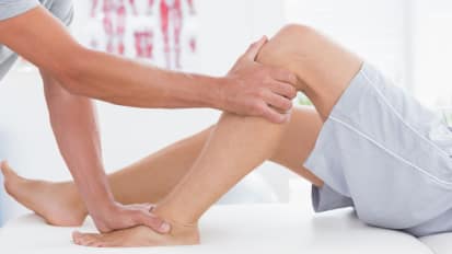
And the ultrasound piece of the visit required the children to lay on their stomachs or to lay prone. And as you can see, this is actually one of our children with cerebral palsy who's laying on our exam table. When they lay prone, we can then get access to the gastroc muscle.
Now, we chose this muscle specifically because it's one of the primary targets-- it's one of the primary muscles that we inject with botulinum toxin. It's also a very important muscle for locomotion and one that's prone to shortening. We have a lot of difficulty with maintaining length of this muscle. So it's a very important muscle from the standpoint of studying cerebral palsy.
So we measure-- so we have them placed on their stomachs and we have them relax and we measure their relaxation ability by using surface EMG, a fairly gross measurement. It's something that we can easily do in the clinic and the kids responded to very well. Here you can see our ground electrode from our surface EMG and then we place two other electrodes over the gastrocnemius muscle itself.
We then passively position the foot in differing degrees of range of motion. So starting off with the muscle in slack and plantar flexion and then increasing to 10 degrees of dorsiflexion, so really putting that muscle on stretch. And we measured four positions total. We did this on each leg and we did this three times.
So what do we get when we do these measurements with the ultrasound? First of all, I'm going to show you my very crude attempt-- oops-- at drawing what we're doing.
So if you could imagine if somebody is-- yourself, for instance, sitting there-- if you think about this as being your leg and this as being your foot, if you point your toes down, you're in plantar flexion. If your foot is flat on the floor, then you're in 0 degrees of plantar flexion. And then if you bring your toes up off the floor, you're in some dorsiflexion. So that's what I'm referring to when I'm referring to the positioning that we did.
When we did our elastography techniques, specifically the SSI, we get a B-mode image. It's superimposed upon this B-mode image, this black and white image we get an elastogram. And let me orient you to what you're looking at here. So this very top portion is the muscle and fascia. Then there's the gastroc muscle, the soleus muscle, and then the big toe flexor below, the flexor hallicus longus.
Our elastogram is superimposed, centered on top of the gastroc muscle. And then within that, we pick a region of interest to be able to do our measurements that excludes these connective tissue areas, because that affects our measurement properties.
The dark-- the color change gives us a visual signal as to what we're measuring, but the machine also does the computations with the shear waves so that we get a quantitative measure. In this child, they're in 20 degrees a plantar flexion here. We see dark blue. That means the tissue is softer, as opposed to-- coming here-- to 10 degrees of dorsiflexion, where the tissue's becoming a lighter green color, which means that we're putting the tissue in more stretch. It's getting stiffer.
So when we look at the kids that we recruited to this study, we had 20 typically developing children, and so far we have 13 children with cerebral palsy enrolled in this study. We compared their demographics and some important measurements with regards to what we're looking at in the calf muscle.
Overall, the age-- and this is in months-- so the age and the body mass index was not significantly different between these two groups. However, the children with cerebral palsy tended to be a little bit younger and tended to be thinner. And the thinner is not surprising.
With regards to calf circumference, they tended to have smaller calf circumference. In fact, this was statistically significant. And their dorsiflexion tended to be less than the typically developing children, though was only statistically significant on the left side.
Now these differences are expected. This is what we see clinically. This is well-documented in the literature. This is not surprising that we are seeing these differences. And below, I'm going to introduce you to some terminology that we use commonly in cerebral palsy, and it's a way of classifying the severity of the movement disorder.
First is GMFCS level which is Gross Motor Functional Classification System. How well do these children walk? A level one is a child who walks really well. Level five is a child who's completely dependent, can't walk, needs somebody else to assist them with all of their cares.
For our study, we were able to enrol children who were levels one to three, with three being a child who uses a walker and may use a wheelchair for longer distances, or uses a wheelchair primarily but is able to self-propel.
When it comes to CP type, we like to classify them based on how many limbs are involved and what limbs are involved. And for this study, we included all children, but were able to enrol children with hemiplegic cerebral palsy, so involving just one side of their body. Some of the children had just their legs involved. That's the diplegic cerebral palsy. And other children had three limbs involved, or triplegic cerebral palsy.
So when we did our measurements, first of all, we looked at our typically developing children. And so to orient you to this graph looking at passive stiffness, on the y-axis here is shear modulus, which is a measure of elasticity, which I kind of crudely call stiffness. Some of the people who do biomechanics may be kind of cringing a little bit in the audience as I'm talking about it in this manner. But it's easier to visualize.
On the bottom or in the x-axis is foot position. So 20 degrees of plantar flexion is when we expect that the gastroc muscle is on slack All the way to 10 degrees of dorsiflexion, when we expect it to be stretched. And as we would expect, our stiffness values increase. This muscle gets stiffer as we stretch it.
And we also see that between right and left sides, the measurements that we get are very, very similar. So similar that they look fairly identical. There was, in fact, no significant difference between the right and left side measurements. But at each foot position, when we compared each foot position to the other foot position, there was a statistically significant difference between those two foot positions, meaning that at 20 degrees plantar flexion, it was less stiff than at 10 degrees plantar flexion, and this was significant.
Another thing to notice is our standard deviations. That's these lines and whiskers. The standard deviation is quite small here when the tissue is softer, but it increases as the tissue gets stiffer. Some of this is due to the measurement capability of the machine itself. As you can imagine, as we're getting stiffer, these shear waves that are traveling through the tissue are traveling faster. It gets harder for the machine to be able to detect these shear waves.
And so we have some inherent variability in the measurement there. There's also a little bit of variability in the measurement just between children. One child's measurement may not be the same as the other child's, like we see when doing any type of study that involves anything living.
Another thing that we looked at was the intraclass correlation coefficient. So when we did those three repeated measures on each leg, did those measures-- or, were those measures similar to each other when we repeated them? And for each foot position we tested, we looked at this. And overall we found that, yes, we had good to excellent results and we looked at these repeated measures within each child.
So now, what happens when we add in a group with cerebral palsy? And how does this compare to our typically developing children? The graphs are set up the same as they were before, with our stiffness measurement being on the y-axis and our foot position being on the x. You'll notice that the x-axis, we don't have 10 degrees plantar flexion here. This had to be eliminated as many of our children with cerebral palsy could not even get into that position to be able to be measured.
What we note-- what you note, and it's pretty obvious here, is that there is a big difference between that typically developing children and the children with cerebral palsy, both on the left and the right sides. And this was, in fact, statistically significant at both foot positions.
The other thing to notice is that the variability appears to increase. So our standard deviation is much larger for our children with cerebral palsy as compared to even our typically developing children. So there's a lot of variability between kids when we're doing these measurements. Despite this variability, we were still able to detect a difference, however.
So what are we able to conclude from this first part of this study? So we're able to directly measure passive stiffness of muscles in children with good to excellent reliability within individuals. Passive muscle stiffness of individual muscles is greater in children with CP as compared to our typically developing children.
And lastly, we were able to find that there is a lot of variation in the measurements between our typically developing children and our children with cerebral palsy. And that's going to affect some of our data coming up here.
So now we're going to look at how botulinum toxin A injections affect our passive muscle properties. Now, for this, we had nine children who completed all three visits. So this is looking at nine children. I took out the standard deviations here, but have placed them in this graph and I have to graphs. They're demonstrating the same thing but in a little different way in order to try and capture this.
And one of the things I'm going to point out is that our y-axis is different here. We have a passive stiffness ratio. And what we did, in order to account for some of that between child variability, was we normalized each child to their own baseline measurement value.
And so down here at 20 degrees plantar flexion, you can see this value one. So this was-- our baseline value for each child was their value when-- their stiffness of their muscle when it was on slack before we did the botulinum toxin injections.
And so before we did the injections, you can see here this blue line, we see the stiffness increasing as we go from 20 to 10 to zero degrees plantar flexion one month after. And this is a mantra in the literature, that we tend to see a peaking of the injections one month after.
And some folks argue that the injections wear off somewhere between three to six months afterwards. And there's no clear cut evidence for which direction this goes. What I can say is it certainly appears that by six months time, we're seeing a loss of effect of those botulinum toxin injections.
And you could see this again here when we're looking at it, specifically at each foot position, comparing it with the bar graphs. The before is the gray. The one month is the brown. And the green is the three months. And overall, when we get to the one month, we see a decrease in that passive stiffness.
And in fact, when we did repeated measures testing, we were able to detect that there was an effective time, meaning the before, one month, three month, and an effect of foot position on our passive stiffness measurements. But because we don't have enough-- we only have nine children, we don't have enough power to dig down into this further to see exactly where these differences appear.
So what can I tell you from all of this? So what I can tell you is that we are able to reliably and noninvasive measure passive muscle properties in children. And specifically, it's reliable within a child.
Passive muscle properties do differ between children with and without spastic cerebral palsy. And passive muscle properties change after botulinum toxin injections. . But this change appears to be transient. Specifically, it appears to be wearing off by time we hit that three month mark.
So this also led me to think about things a bit differently. How are we going to optimize muscle properties in children with CP? How do we make their muscle properties more like the typically developing children, to help them with function in normal growth?
It seems that we really need to actually intervene before we get to this point where we're seeing this vicious cycle of the active muscle stiffness and the passive muscle stiffness. CP really is chronic, and by the time we see a difference or able to detect this in physical exam, the condition's really been present for quite some time.
And so the muscle stiffness that we are trying to measure is truly a dysfunctional adaptation to this chronic over-activation. So it gets to the point of, what's causing this chronic over-activation? What's causing this spasticity that's leading us down this cycle?
So I'm going to get into this through two kids that I follow in clinic routinely. And these are MRIs of the brain of each of these children. The first one is a T2 weighted MRI the second one is a T1 weighted MRI. The second child didn't have a T2 MRI to be able to compare, but I think you can get to the point of where I'm going with these in just a minute.
So first of all, cerebral palsy-- cerebral. Cerebral palsy is defined as a non-progressive disturbance of the fetal or infant brain. That's key to this. And this results in the difficulties with muscle control, creating impairments in posture and movement. This is the definition that we've been living by for years.
Now, infants, a full gestation for an infant is 40 weeks. Both of these children were short of that by some time. In fact, they were borderline extremely premature. In this first child, born at 27 weeks gestation, was able to sit independently by one year of age. For those of you who may not recall, six months is normal for sitting. And this child was able to walk independently by 18 months of age, which is a little bit delayed. 12 months is typically normal.
Child number two, 29 weeks gestation, but able to sit independently at seven months, so really close to being normal, and walked independently at 21 months of age. Fairly similar development with regards to their walking abilities.
But when we look at these MRIs, not only is the type of MRI that I'm showing you different but the brain's imaging in this is distinctly different. In fact, child number one has a normal MRI. We could not find an abnormality in this MRI to explain the difference in this child's development. But this child was born premature. In fact, I went and had this child undergo an MRI of their spine. There was no differences there.
When we look at child number two, this. Child has large ventricles. You wouldn't even want your 90-year-old grandmother to have ventricles that look like this. These are large. And the reason these ventricles are large is because this child has atrophy of their brain related to their prematurity. They have more pronounced sulci and gyri. Their brain is not beefy and as robust as child number one.
So we have these two very different appearing brains. But these children have very similar phenotypes. Their development is relatively similar. Their abilities are relatively similar. So if cerebral palsy is truly all due to a brain abnormality, why can't we predict what their phenotype is going to look like, what their functional impairment's going to be? And we can't even diagnose it based on the brain imaging. A normal brain does not exclude cerebral palsy.
So what else is contributing to this functional impairment? That's the million dollar question. So I'm going to go back to this and get to that, what we think may be contributing to this.
So, again, Botox, selective dorsal rhizotomy, we use these to tackle our active muscle stiffness. Botulinum toxin, that neurotoxin that we inject in the muscle, and it acts, blocking the ability of the nerve to talk to the muscle.
Selective dorsal rhizotomy, however, something that blocks input to the hyper-active motor neurons, to over-excitable motor neurons. And basically, motor neurons that respond to this low level stimulation are hyper-excitable or spastic and we're trying to reduce their input through our selective dorsal rhizotomy.
The stimulation technique that we use for the surgical procedure is very similar to something called an H-reflex, which is done routinely in our EMG lab, to measure for motor neuron excitability in folks with suspected motor neuron disorders.
And as we were talking about this, it suddenly came clear that, why can't we use something like this for early diagnosis and early detection of motor neuron hyper-excitability? This would also provide an opportunity to intervene before folks even show the signs of the phenotype of cerebral palsy, and perhaps impact how their development is going to be down the road and reduce the need for things like our botulinum toxin, selective dorsal rhizotomies, and our other interventions.
So let's think a little differently about spastic cerebral palsy. And let's think about it a bit as a motor neuron disorder, but specifically, developmental motor neuron disorder.
So this is distinctly different from ALS. So if we think about ALS, that's a degenerative motor neuron condition. This is not a degenerative condition. This is a developmental difference of the motor neurons. So we believe cerebral palsy is a disorder due to difference of motor neurons because of an insult at a critical period in neuro development.
So critical to this conceptual framework is the transition of glycine and GABA. And this may be something that's new to many of you out there. I know at the time that I've been working on this, it's something that was new to me. So glycine and GABA are actually excitatory early in development and they become inhibitory by time we reach maturation. And this is normally how motor neuron pruning occurs.
In animal studies, it's been shown that decreased motor neuron activity during the embryonic period and early postnatal period results in a greater number of motor neurons. So if we have decreased input either through blocking some of the excitatory neurotransmitters or by blocking muscle activity itself, motor neurons can't prune. We end up with too many motor neurons.
So what happens in our kids that are born premature? Well, they may actually be taking two hits. One, they may have some effect for the glycine, GABA input to the motor neurons due to disruption of their central nervous system. But the other is they may have some decreased movement due to sedation. Often, these babies are very fragile when they're born.
There's a limited window of ideal oxygenation and ideal blood pressure that we're trying to maintain primarily to protect the brain. But what if what we're doing to protect the brain is actually causing a greater impact on those motor neurons and resulting in the phenotype that we're seeing in cerebral palsy?
So if that's the case, what we would expect is we'd see a knock out of the glycine and GABA, see this excitatory input early on. But it's not as much excitation as we would see in the normal developing infant. And so in the normal developing infant, the motor neuron pool prunes appropriately.
In a child, in an infant who goes on to develop CP, they may have this, plus the reduced muscle activity from being sedated very early on. So they have this larger motor neuron pool.
Now we move forward, that CNS disruption that they have is permanent. So the glycine and GABA are still knocked out. They have this larger motor neuron pool, but now they have no ability to inhibit this. So once they start doing an activity or their muscles start activating, there's no way to quiet this down. This is what we see in that reflex loop of spasticity. We don't see that in folks who don't have spasticity because they have that normal inhibition there to shut that down.
So our hypothesis is that spastic cerebral palsy results from an increased number of motor neurons due to that reduced motor neuron pruning early in life and increased motor neuron excitability, which results in spasticity and impaired physical function.
How are we going to explore this novel paradigm with cerebral palsy? So we're going to do it two ways, both through looking at individuals who have cerebral palsy but also in an animal model. And specifically, we're going to assess motor neuron abnormalities in individuals who have cerebral palsy. We're going to do this through some methods that we know that have been used for quite some time but have not been used to systematically explore this in this condition.
So first of all, what we're proposing to do is to work with individuals with CP and do an H-reflex, which tests for the excitability of the motor neuron. And then look at motor unit number estimate, which is an indirect way of counting motor neurons.
Now, for those of you who work with animals and maybe do some of this yourself, you know that to truly count a motor neuron, you need to be able to stain that motor neuron. You need to be able to section the spinal cord, and then go in and physically count them. We can't do those in people. So what we're going to do is actually estimate the motor unit number.
Now, starting with the H-reflex, we're looking at doing a novel technique for doing the H-reflex. Dr. Litchy has been spending some time in his lab trying to get an H-reflex response from the fibular nerve. This is a nerve to the anterior tibial muscle. And in individuals who have a normal central nervous system, he has not been able to get this response.
Now, we can get it in other muscles. So you can get it in the tibial nerve to the gastroc muscle or in the ulnar nerve, but it can't and the fibular. And so we have an individual with spastic cerebral palsy and we tried to do this in that person.
And lo and behold, we were able to get a fibular response. So part of our research is going to be, can we consistently get this response in individuals who have disinhibited central nervous system, who have a hyper-excitable motor neuron, and be able to use this as a yes/no response? If not, we still have the classic response to be able to use, and I'll show you that in a minute as to how we can use that.
Our motor unit number estimate is a little bit trickier. And this requires, again, electrophysiologic testing. Basically, you stimulate at sub threshold and then you get individual responses. So this is in microvolts, and we'll get individual responses which are individual motor units that are responding to your stimulation. And you very gradually increase this.
And in fact, this group did this up to approximately 440 microvolts and they had 11 increments. So when you do the math, that gives you about 40 microvolts for each response of the motor neuron.
They then went on and did supra threshold stimulation, so basically got the whole muscle to respond. And got-- eight microvolts was the total response for the whole muscle. So you take the eight microvolts, you divide it by the 40-- or, the eight millivolts divided by the 40 microvolts and you get about 200 motor neurons.
And we'll be able to do this in our individuals with cerebral palsy, which is not something that's been done before, to give us an estimate. Do they truly have too many motor neurons?
We can also do this same thing in mice and we are going to do this in a spastic mouse model. Now, the reason we chose this mouse model is it has a glycine receptor mutation. So it has what mimics what we believe is going on in spastic cerebral palsy. And it's not been used to explore spastic cerebral palsy in this way. It's been used to look at Botox. It's been used to look at contractures, but not at the motor neurons specifically.
Now, there is other animal models out there where they try to inflict the brain injury that happens in CP, but those animal models don't develop spasticity, which is a key component of cerebral palsy. So we really need to get to the heart of the spasticity.
So in the mouse model, we're going to do the H-reflex, similar to what we're doing in our human with the fibular nerve. But if that does not work, our back up is to do the typical H-reflex testing. And this is an example of this. So doing the tibial nerve, if you do an H-reflex test and you try it at different levels of stimulation, so different frequencies of stimulation, as your frequency of stimulation increases, naturally, the reflex decrements. It dies out. It becomes inhibited. In an individual-- in this case, a rat-- who doesn't have this disinhibition, you don't get that decrement. And so you can compare the decremental response to be to look, is this hyper-excitable or not?
Lastly, we're going to be looking at motor neurons through direct motor neuron labeling. We can do this through use of cholera toxin B, which is used in-- cholera toxin subunit B which is used regularly in Dr. Sieck's lab, who we'll be working with for this. And through this, we can label the motor neurons specifically, the ones for muscles that we are interested in, and be able not only to count motor neurons but also can use this to estimate motor neuron size, too.
So future directions. When all of this pans out, things that we planned to do with this are to use our animal model that we're developing here to clarify what's that critical window for the neuroplasticity of the motor neuron? Where do we need to intervene to really get the biggest bang for our buck?
And then look at animal strategies-- or, animal studies on potential regenerative rehabilitation strategies. How do we help with function? And how do we help with function before there's even a problem? How do we prevent a functional problem?
One way we can do this is looking at sedation. How the sedation affect these animal models? How does this affect the motor neuron pruning? If we do muscle stimulation, low level, different levels of stimulation, can we affect motor neuron pruning? And can we use medications to impact that? Can we use things like GABA agonists to affect motor neuron pruning. Then the long term goal is to take the techniques that work the best and are safe into clinical trials, specifically, clinical trials with our infants, which is, clearly, sometime down the road.
So I'd like to thank individuals who've been involved in my research, and specifically Dr. An and Dr. Sieck who have been instrumental as mentors and getting me to the place that I am today. Dr. Greenleaf and his lab for introducing me to the ultrasound technique and supporting me in that. And the clinical colleagues I have that have been instrumental in helping me with recruitment and carrying out the studies. Thank you.
[APPLAUSE]
VICTOR MONTORI: 4:41, so there's gong to be a time for questions. Hang in there. [INAUDIBLE]
AUDIENCE: Hi, Joline. Thanks to a very exciting talk. I was wondering, if your hypothesis is correct about the neurotransmitters and hyper-excitability-- which is a great hypothesis, I think, by the way-- do antagonists of excitatory neurotransmitters in cerebral palsy children have any effect on spasticity?
JOLINE BRANDENBURG: So at this point, we don't use antagonists. We typically use agonists of inhibitory neurotransmitters. So one specific medication that we use is baclofen, which is a GABA agonist. And that seems to help with the spasticity.
The problem with baclofen at least, in our kids, is that there's a lot of difficulty with sedation with it. So I'm not sure about using that. But it's something that we can look at, and look at specifically in the infants. It may have a paradoxical response because the neurotransmitters are different in that early stage of development. And so using something like what's traditionally thought of as an inhibitory neurotransmitter, GABA, early in development, it's actually excitatory.
VICTOR MONTORI: Another question? While people are thinking, it's interesting as we go through the professional pathway and we start thinking about these ideas when we're thinking about the different [INAUDIBLE], there's some different therapies that result from thinking differently about the disease, one is supposed to be thinking very quickly about intellectual property, patents, and that sort of thing. Can you tell us about your journey in that road?
JOLINE BRANDENBURG: So unfortunately, I have no patents or intellectual property. But our ultrasound lab has quite a bit. And the elastography technique we are using currently is not property of our ultrasound lab, but they are developing techniques to optimize. And so they're working with industry with regards to that. But at this point, I don't have anything that's in that realm.
VICTOR MONTORI: There's another question.
AUDIENCE: I have another question if I have an opportunity. In the beginning of your talk, it was really fascinating to me that you were talking about one of the reasons for spasticity in muscles is build up of collagen. And I know in our spinal cord injury research that we sometimes use enzymes that degrade those kind of collagens and that kind of thing to promote repair. And I was wondering, since therapeutically, Botox is already injected into muscle if you can inject collagenase or another enzyme that might loosen up the muscle fibers and allow it to relax enough to do the exercise, et cetera?
JOLINE BRANDENBURG: That's a very interesting thought. I'm not aware of using the collagenases, although I think there's something they use similar for things like Dupuytren's contracture. And some of those, collenases, when you inject them, they spread to a lot of other things than what you want it to break down. And so at this point, I think I would be worried that it would not only break down the collagen but also would have a negative detrimental effect on the muscle itself. But that would be a very interesting area to target. Oh, dear.
AUDIENCE: [INAUDIBLE] So I had a question. [INAUDIBLE]
[LAUGHTER]
AUDIENCE: Thanks for the talk, Joline. I have a question specific to your children that had the hemiplegic type of cerebral palsy. Since you looked bilaterally, did you notice a difference in stiffness where maybe there was a global effect, even though one side was the affected side?
And then, secondarily to that, do you think those children, or the differences in limbs that are affected, may help determine if it's a motor neuron issue or the development of the motor neurons, versus a specific brain injury? Because I could see the motor neuron potentially being a global effect, if it's related to the neurotransmitters. You'd think it might be widespread as opposed to focus in one area.
JOLINE BRANDENBURG: That's it. That's a great question. So with regards to question one, we did. We did look at both sides. And that was part of the reason I wanted to look at both sides was to see, is the hemiplegic side truly unaffected in our individuals with cerebral palsy?
And it's-- we have five or six kids in that group and it looks like their muscle stiffness on their unaffected side is slightly greater than children that are typically developing children. But it's definitely less than cerebral palsy. So there is some effect. Whether or not that's a compensatory measure, you know, they have this over-activity on one side. Does that somehow affect the other side? Or is it a compensatory measure because maybe they're toe walking all the time on that side and it's affecting the stiffness of their muscle because of the way they're activating that calf muscle. That part we don't know.
From the motor neuron perspective, . interestingly, folks with-- the kids with hemiplegic cerebral palsy tend to have a focal-- they've had something occur like a stroke, a focal lesion that contributes to their cerebral palsy. And so they may have a combination of that stroke and potentially a motor neuron disorder. But for them, it appears to be more that it is truly the stroke that's interfering with that, though their phenotype may differ, because of the impact of that stroke on their development in their motor neuron development on that side of their body.
VICTOR MONTORI: This is going to be the last question.
AUDIENCE: Thanks, Joline. Spasticity is a common problem in a number of conditions, so stroke you just mentioned, brain trauma, spinal cord injury. Do you think there's a common mechanism that might exist that could be applied across the board?
JOLINE BRANDENBURG: So the common-- so for individuals, and there are individuals who develop spasticity later in life, say, through spinal cord injury, traumatic brain injury, and then those individuals, they've already had the normal motor neuron pruning and they have that intact motor neuron pool. But there may be a difference in those neurotransmitters that's affecting them, affecting their spasticity. So I think the common denominator is looking at that and looking at the neurotransmitters and the impact of the spinal cord injury or the brain injury on that.
VICTOR MONTORI: All right. Please join me in thanking Dr. Brandenburg.
[APPLAUSE]
[SIDE CONVERSATION]
JOLINE BRANDENBURG: I can't statistically prove that they're worse, but clinically it's interesting. When we go back and repeat it, we don't get as great an effect the second time or third time around as we do the first time. And I'm almost wondering if it's [INAUDIBLE] that's that point of interference [INAUDIBLE].
SPEAKER 1: Right.
JOLINE BRANDENBURG: Now, we have this--
SPEAKER 1: Just theory.
JOLINE BRANDENBURG: --which is yeah. And we have this temporary relief from the spasticity. Right now, we have nothing else that is used for that. And so there's been a couple of papers out there looking retrospectively at effective [INAUDIBLE] botulinum toxin [INAUDIBLE] range of motion, [INAUDIBLE] showing that there's no change or no [INAUDIBLE] range of motion [INAUDIBLE] who've had botulinum toxin injections.
SPEAKER 1: [INAUDIBLE]
JOLINE BRANDENBURG: [INAUDIBLE] might be doing something to the [INAUDIBLE] works, which is part of what started me on this. But I can't--
SPEAKER 1: [INAUDIBLE] Sorry. [INAUDIBLE]
JOLINE BRANDENBURG: Sweet. Are you two going to peds?
SPEAKER 2: Yeah, going to do peds. Start neuro in a week.
JOLINE BRANDENBURG: Start neuro in a week. Neuro's tough. Are you on inpatient or outpatient?
SPEAKER 2: I don't know how the schedule works. It's kind of crazy [INAUDIBLE].
JOLINE BRANDENBURG: OK. Where did I put my jump drive? I'm thinking that [INAUDIBLE].
SPEAKER 1: Oh, wow. Thank you.
SPEAKER 2: Thank you.
SPEAKER 1: Yes.
JOLINE BRANDENBURG: Just got to find my jump drive.
SPEAKER 1: Oh Did he give it back to you?
JOLINE BRANDENBURG: I'm not sure. I hardly remember.
[LAUGHTER]
[INAUDIBLE]
SPEAKER 3: Let me tell him that.
JOLINE BRANDENBURG: OK. I don't remember getting it back, but-- you have to go to clinic, dear, right?
SPEAKER 4: Sorry. [INAUDIBLE]
JOLINE BRANDENBURG: You see it?
SPEAKER 4: [INAUDIBLE]
JOLINE BRANDENBURG: No, it's the one that's on the key chain. There it goes. We've seen me use it.
SPEAKER 4: [INAUDIBLE] looks like my car keys?
JOLINE BRANDENBURG: Yeah. [INAUDIBLE]
Yeah. Then you ran it up here. That's right. He might have dropped it [INAUDIBLE].
SPEAKER 4: [INAUDIBLE]
SPEAKER 1: It's not this little Guy?
JOLINE BRANDENBURG: What? Nope.
SPEAKER 1: OK.
JOLINE BRANDENBURG: Oh, wait. Yeah, yeah, yeah, yeah, yeah, yeah. Yes.
[LAUGHTER]
Yep, yep, yep. I would have never seen that. OK.
SPEAKER 4: Found it.
JOLINE BRANDENBURG: [INAUDIBLE]
[INTERPOSING VOICES]
SPEAKER 1: Got it? Perfect. Excellent.
JOLINE BRANDENBURG: Thanks again. Have a great weekend. Yes.
SPEAKER 1: [INAUDIBLE]