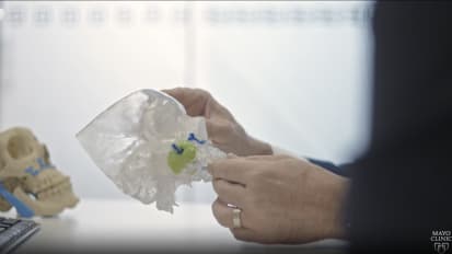
DALE EKBOM: Hello, this is Dale Ekbom. Today we're going to talk about microflap excision of a vocal polyp. There's lots of different polyps, all different shapes and sizes. And one of the keys is to just remove what's abnormal. You're trying to save all the superficial lamina propria if you can. You're trying not to even see the ligament because you want to leave that area of superficial lamina propria over the top. And this is more epithelial work, as well as removing more just the polypoid tissue.
So here's a polyp in the left true vocal fold. Closer up shot, and you're seeing normal tissue and then going right into abnormal. And that's where we began our incision. This is our 70 degree pictures as well. And looking on the other contralateral side, too, to make sure there's no reactive change.
So I start with my suction or a probe and just get a feel for where is this polyp attached to. Often it's on the inferior lip of the true vocal folds. This is a sickle knife that we start with. And you try to have a very sharp sickle. If it's not so sharp, you can start it with the sickle and then finish with an up scissors if you need to.
But here we are extending it. It's bleeding a little bit, which is typical with the blood vessels that feed into these polyps. So I have an epinephrine-soaked pledget here. It's 1 to 10,000 epinephrine. And that stops any sort of oozing in that area. It also can be used a little as a Kittner just to push around and just dissect a little bit of the polypoid contents off of the superficial lamina propria. So there, you're seeing within the microflap right now.
Next, I have my Ossoff microflap elevator. And I push more laterally, but I'm just wanting to just remove polypoid contents. And so then I take this up and down, anterior, posterior, to carefully excise out any of the polyp that's there, leaving a nice, smooth edge deep. I use the graspers just to pull-- just to grasp just the edge of the epithelium there. You see how careful you are in pulling that out. So you can see the inside of the microflap. And then you dissect some of the inside portion up away from the inferior portion of the microflap. And then I make an incision as I've dissected up the polypoid contents so that my inferior flap will lay up against my superior flap.
So that's dissecting out the contents. You can go through with your microflap elevator. Or you can use a scissor. This time, I went through with the elevator. And that works fine.
And then I take my up scissors and just finish the cut, trying to avoid a dog ear. So you'll go right at the corner. And you connect it. And that will prevent you from having to re-excise. This anterior portion is always difficult. You have to really, again, go at an angle here. And that typically prevents excess tissue.
And if there is any residual, you can use a laser to remove that if you can't get it with the scissors. And then I push up the lower flap so it connects to the superior one. And it looks great with a 0 degree scope. And we also look at it with the 70 degree scope.
If there's a little gap, it's not bad because you've gotten that lower flat very close. And it typically heals really well. There's excellent re-epithelialization typically. But you do want to get those flaps as close as you can together. So that's how we do a microflap procedure. Thanks for listening.