Chapters
Transcript
SPEAKER 1: Hybrid pancoast tumor resection, by the division of thoracic surgery, Mayo Clinic, Rochester. In 1932 the radiologist Henry Pancoast first described a superior pulmonary sulcus tumor as a carcinoma developing in the apex of the chest. A newer definition of a Pancoast tumor includes, according to [INAUDIBLE] and colleagues, a lung cancer arising in the apex of the lung that involves structures of the apical chest wall.
Involvement of the chest wall only at the level of the second rib or lower should not be considered to meet the criteria for involvement of the apex of the chest, and the chest wall involvement may be limited to invasion of the parietal plura, or may extend deeper to involve the periosteum or the bone of the upper ribs or apical vertebral bodies. Or it may include invasion of the subclavian vessels, the nerve roots of the brachial plexus, or the stellite ganglion.
Traditional approaches for Pancoast tumors involve the posterolateral Shaw Paulson approach, a transclavicular Dartevelle approach, the trans-sternal Masaoka approach, and a hemi-clamshell approach.
Our case is of a 64-year-old woman who presented with a 30 pack year smoking history. She had moderate aortic regurgitation and presented with a tumor in the right upper lobe of her lung. The tumor measured 10 centimeters in maximum diameter, was adenocarcinoma by biopsy, and was TTF-1 positive by immunohisto chemistry staining.
And she was found to have the involvement of the T1 nerve root. And a bronchial ultrasound was performed, and all of the mediastinal lymph nodes that were sampled were negative for cancer. An MRI was performed, which also showed involvement of the T1 nerve root. The initial chest X-ray can be seen in the image to the left, with the tumor in the apex of the right upper lobe of the lung.
After neoadjuvant therapy, considerable shrinkage of the tumor occurred, with reduction of the volume by almost 50%, an involvement clearly showing re-expansion of the superior segment of the right lower lobe, with no more compressive atelectasis, and the tumor limited to the apex of the right upper lobe of the lung, extending into the T1 nerve root.
Additional imaging by CT again shows the reduction in the size of the tumor after neoadjuvant therapy. The MRI clearly showed involvement of the T1 nerve root, however preoperative 3D printing in adjunctive use with MRI and CT scan aided us in guiding our planned resection, and clearly demonstrated involvement of the T1 nerve root.
Traditional VATS ports were used, with an anterior axillary line fourth to fifth intercostal space utility incision, two additional thoracoscopic ports placed, one in the anterior axillary line in the fifth to sixth intercostal space, one in the posterior axillary line in the fifth to sixth intercostal space, and then a limited posterior access incision was used to access the posterior detachment area of the first and second ribs through an open approach, but limiting our incision, no longer cutting the latissimus muscle, no longer cutting the serratus muscle, and no longer spreading the ribs. The right upper lobe was removed with video camera surveillance, and hyler vessel and bronchial division with staplers.
A time out procedure was performed at the beginning of every case, where the entire team pauses, and a briefing is performed. Following this, we review planning with the 3D model. The 3D model showed us clearly which ribs would have to be removed to resect the tumor. The tumor is black. The brachial plexus is the lightest gray color. The right-sided subclavian artery is the intermediate gray color, and the subclavian vein is the darker gray color. We met with the orthopedic surgical team to plan our resection.
We begin our surgery by placing the two camera ports and the anterior utility incision, only after positioning the patient in the left lateral decubitus position. The posterior plural dissection is performed first. By gently spreading, we can delineate the edges of the right upper lobe bronchus, and see the Azagus vein quite clearly. The attachments of the right upper lobe apex tumor can be seen to the chest wall. We then begin by flipping the lung posteriorly, and beginning an anterior to posterior dissection.
A small peanut, or small sponge can be used to dissect the tissue away from the phrenic nerve, spreading all the lymphatic tissue away from the superior pulmonary vein branches. Care taken to preserve the right middle lobe pulmonary vein branches, such that they are not inadvertently stapled and divided. Preservation of the right-sided phrenic nerve is made throughout the surgery by identifying it with the camera. Removal of the lymph node anterior to this phrenic nerve will have to be performed as part of the standard lymph node dissection.
We encircle, staple, and divide the superior pulmonary vein branches going to the right upper lobe and then encircle, staple, and divide the right upper lobe truncus anterior branches. The fissure between the right upper lobe and the right middle lobe, and the right lower lobe, is then taken to facilitate exposure of the poster ascending pulmonary artery branches coming off the posterior aspect of the pulmonary artery after we have easily divided that portion of the truncus anterior. By taking more of the fissure, this enhances our angle and manipulation of the stapler such that we can now clearly see, in the center of the screen, the posterior ascending pulmonary artery. The stapler, a tan load curved tipped, is used to pass around the posterior ascending pulmonary artery by putting our camera in the more posterior approach, and the stapler and the more anterior inferior port.
A small portion of the posterior ascending pulmonary artery has not been divided by our stapler. And it is important to remember, when removing your stapler, not to simply pull it back quickly, as you could potentially disrupt the pulmonary artery. We use scissors to divide this remaining area, and then gently pass a zero silk suture circumferentially around the right upper lobe bronchus. The lymph nodes are swept up along the edge of the bronchus, and a purple load endo-GIA stapler is used to staple and divide the right upper lobe bronchus. After the right upper lobe bronchus has been removed, we then begin our portion of the chest wall resection. A hooked cautery used to divide the tissue circumferentially around the attachment of the apex of the chest wall. And a ligature devices is used to facilitate the dissection of the tissue, the intercostal muscle, superior and inferior to the second rib. This portion of the surgery entails division of the anterior aspect of the right second rib, which is now being divided with the [INAUDIBLE].
By sequentially dividing this anterior portion of the right second rib, we can use the ligature device to control bleeding from the intercostal artery. Cautery can be directly applied to achieve hemostasis at the intercostal artery. Once the anterior aspect of the right second rib has been completely divided, we then begin our posterior approach by using the [INAUDIBLE] to additionally divide the anterior margin of the second rib, we can completely mobilize it from its attachments to the chest wall.
During different portions of the surgery, we made different passages to divide that anterior attachment of the second rib. And during some portions of these surgeries, we had already created our posterior incision. In a few moments, we'll show you our post here accessory incision, which is being created now. We use Bovie electrocautery with a limited posterior access incision-- no rib spreading-- to divide the trapezius and the rhomboid muscles directly. We are taking care not to disrupt any of the scapular vessels. We are also making sure that we gently spread the perispinal muscles away from the anterior attachments, such that mobilization and exposure of the entire right apex of the chest wall can be achieved.
Once the posterior access incision has been made, we then began to divide the posterior scalene muscle, which is attached to the right posterior aspect of the second rib. This posterior is scalene muscle is divided, and then attention is directed towards dividing the anterior and middle scalene muscles to provide access to the lateral aspect of the first rib.
A periosteal rib elevator is used to elevate the tissue off of the posterior aspect of the right second rib. A DeBakey forcep is used to retract the perispinal vessel away from the posterior aspect of the right second rib, and a [INAUDIBLE] again is used to divide this portion of the rib. Bovie electrocautery is used to divide the anterior and medial attachments of the perispinus muscle, retracting it posteriorly such that we can easily see the articulation point between the apex of the chest, first rib to the transverse process, and second rib. By using a finger to dissect through this tissue, we can then easily see the posture articulation of the second and first ribs against this lateral aspect of the spine. It is with the Cobb elevator that we then use to disarticulate these portions of the rib heads from their lateral attachments.
Additional Bovie electrocautery is used for hemostasis. We are careful to detach the posterior attachment of the scalene muscle up to the second rib, such that we can expose what will eventually be clearly seen as the brachial plexus, the subclavian artery, and the subclavian vein. As the posterior scalene muscle is elevated, we then are provided with excellent exposure of the thoracic outlet structures.
At this point a joint dissection is performed between cardiothoracic surgery and orthopedic surgery. The thoracic surgeon is responsible for detaching those portions of the rib that are directly adjacent to the tumor, and the orthopedic surgeon is carefully navigating the disarticulation from the transverse process of the vertebral body. Using a rongeur with a narrow tip, we are then able to complete the bony detachment of the posterior aspect of the right second rib. Now that this right second rib has been released from its posterior attachment, it is now being mobilized with the Cobb elevator.
This mobilization enhances visualization both on the outside view as well, as on the inside view. The scapular elevator facilitates completing exposure as we detach the posterior aspect of the scalene muscle to the inferior posterior aspect of the ribs. We detach the intercostal muscles from the superior aspect of the right third rib, and now our dissection of the inferior portion of the chest wall is complete.
Hemostasis in this area is very important, and we take special care not to place any gel foam or hemostasis agents that may be entrained into the epidural space, in the event of a spinal leak. Now that we've completed the posterior attachment of the second rib, we're turning our attention towards disarticulating the posterior aspect of the first rib. During this portion of the surgery, we are now transacting the T1 nerve root.
Because the T1 nerve root was invading into the tumor, it was impossible to separate or spare that T1 nerve root in the process of removing the right upper lobe tumor. By detaching the posterior aspect of the first rib, dividing the T1 nerve root, we have completed the posterior attachment portion of the surgery, as we are detaching the chest wall tumor from its attachments posteriorally. We will then turn our attention towards transacting that superior aspect, dividing the intercostal muscle along the apex of the chest.
The scalene muscle division is then performed with a thoracoscopic view. Taking the scalene muscle with a bipolar energy device allows us to more completely and carefully visualize what we're dividing. At the same time, we can easily use a finger from the external approach to palpate those structures that are lying underneath the intercostal muscle. At this point in time, we're now dividing the middle scalene muscle, which is interposed between the brachial plexus and the subclavian artery.
Eventually, we will then divide superior to the right second rib attaching to the first rib, the anterior scalene muscle. Once the anterior scalene muscle is divided, one can then easily visualize the subclavian vein. Using a right angle clamp to encircle the anterior scalene muscle, we are then more easily able to use electrocautery to divide the muscular attachment, interposed between the subclavian artery and vein.
Pulstations of the subclavian artery can be seen during this dissection, and visualization of the apex of the chest also provides an excellent view of the brachial plexus. We are now dividing the intercostal muscle between the second and the first rib and taking the periosteum off of the anterior aspect of the first rib. This anterior division of the first rib from its medial attachment can be done with a small burr.
The burr has the advantage of not dividing any tissue and only dividing bone. Being selective for bone, and deselecting for tissue, we limit our chances of damaging any underlying tissue, such as the subclavian vein, which is lying directly posterior to our anterior aspect, and superior aspect of the first rib on the right. The burr is then used to completely transact that portion of the medial attachment of the right first rib. As the right first rib is divided more completely, you can then see the apex of the chest wall becomes much more mobile. And we are then able to manipulate the tumor in a slightly more advantageous way, such that we can complete our resection.
The Cobb elevator is then used to disarticulate the anterior medial aspect of the right first rib from its medial attachment. A [INAUDIBLE] is then used to take a small additional portion of the rib that is left remaining, taking care not to get too close to the right subclavian vein. Taking it small in this manner, bit by bit, we decrease our chances of inadvertently and encompassing some of the remaining tissue that may be behind this, such as the vein or, more laterally, the artery.
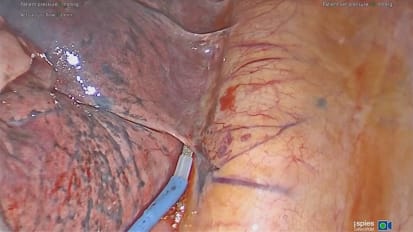
The thoracoscopic visualization of this portion of the surgery is excellent and provides an outstanding arena from which to teach residents during the surgery, when often only a single surgeon can view what's being performed from the outside of the chest. We then complete detachment of the mediastinal tumor attachments, place the tumor into an endo catch bag, and remove it from our open incision. Now that the tumor has been removed, the apex of the chest can be clearly seen.
The retractor was just adjacent to the T1 nerve root. The subclavian artery can be seen pulsating. We are now pointing to the scalene muscles that are interposed just posteriorly to the brachial plexus and the subclavian artery.
The posterior scalene muscles, the middle scalene muscles, and the anterior scalene muscles are all being pointed to. The subclavian vein is just to the right of the pointer. The brachial plexus is at the tip of the pointer. And adjacent to that is the subclavian artery. The pointer is now on the subclavian vein.
Once we have removed the tumor from the chest, it is sent for pathologic evaluation. The margins, both the bronchial and pulmonary artery and chest wall margins, are all found to be negative. The tumor was completely removed, all margins negative, all lymph nodes negative, a complete mediastinal lymph node dissection was performed. The patient was discharged from the hospital on post-operative day number five. She was taking Tylenol only for pain and has returned for follow-up with good re-expansion of the right middle and lower lobes, and no residual problems. Thank you for your attention.
Shanda Blackmon, M.D., M.P.H., professor of surgery and thoracic surgeon at Mayo Clinic in Rochester, Minnesota, shares a case presentation on hybrid Pancoast tumor resection.
3D printing is becoming more widely used in medicine, particularly in preparing for complex cases. The technology facilitates multidisciplinary preoperative surgical planning, assists in the navigation of complex or obscured anatomical relationships, and allows surgeons to rehearse surgical approaches on models prior to taking the patient to the operating room.
This technique may enhance a more rapid progression to more advanced surgery by allowing surgeons to practice and think about their cases with life-sized models that are a virtual simulation of the case they have to plan. The model proved to be invaluable in this novel, first-reported, hybrid minimally invasive approach to a large superior sulcus tumor.
Related Videos