MATTHEW J. BAK: My name is Matthew Bak. I'm Assistant Professor in the department of otolaryngology here at Eastern Virginia Medical School. My specialty is, specifically, head and neck cancer and microvascular reconstruction. And today, I'll be going through a complex reconstructive case, where I use free tissue transfer using a patient's fibular bone and skin from the lateral aspect of their leg to reconstruct an ablative defect after my partner, Dr. Karakla, removed an oral cavity cancer.
Part of our surgical planning for these cases, ahead of time, we will obtain a fine-cut CT scan of the patient's mandible. And then we're able to use that data, import it into a computer program, where we're able to use rapid prototyping and 3D printing to allow us to plan ahead of time and design a reconstruction that's patient-specific and is based on a 3D model that we created of the patient's native mandible and the reconstruction that we plan. So we work with our colleagues in the medical device industry to do this.
So you can see here, this is the planned outcome, and moving forward, we'll go through each step that leads us to this outcome. And this is done approximately two weeks before the planned operation. Initially, resecting surgeon will specify where the cuts will be on the patient's mandible, indicating how much jawbone he is going to resect during the cancer part of the operation. This is noted here in red.
Then we remove that segment and we replace it with fibular bone. And this can either be the patient-specific fibular data or generic fibular data that the company has. And then were able to design a reconstruction plate to span that gap and allow us to bring the fibular bone up to reconstruct the mandibular arch. This plate is made out of titanium. It is not reactive.
And you can see here, the computer design also allows us to avoid injuring tooth roots and the inferior alveolar nerve. In this segment here, these are the cutting guides that allows the plate to fit exactly the way we want it. Then there is also a cutting jig that we'll get for the fibular bone that allows us to cut the bone exactly the right size and angles that we need it to fit the defect. This is especially important when we have multiple segments of fibular bone that we need to perform in the reconstruction, as the defect isn't always a straight line. If you take out the anterior portion of the mandible, the fibular bone needs to be cut in pieces to allow to reconstruct that arch.
This is during the harvest of the fibular flap. We're basically making the posterior cut of the skin paddle here, and it looks like most of the harvest of the fibular bone's already been performed here. The fibular bone has already been cut from the patient. Now we're freeing up from its muscle attachments. You can see the skin paddle that's associated with the bone.
The vascular supply to the bone is the fibular artery, and the pedicle, the artery runs along the medial aspect of the bone and will send cutaneous perforators, or blood vessels, that supply blood to the skin along the posterior aspect of the fibular bone. So we're able to harvest a composite flap of both bone and skin and even some muscle.
You can see the skin paddle now, closely associated with the fibular bone. The dot of the purple marker on the skin paddle marked the blood vessel that supplying the skin, and the skin paddle is centered around that blood vessel that supplies it. And these are the pedicle vessel, the fibular artery, and the two [INAUDIBLE] that are being dissected [INAUDIBLE].
Here, you can see the 3D printed model of the patient's mandible in the planned reconstruction. This helps us intraoperatively with placement of the reconstructed plate and also planning the surgical resection. It can come in handy if the ablation or the cancer part of the operation changes and that the defect becomes larger.
While we harvest the fibular flap, we have a tourniquet on the leg to limit the amount of bleeding. So once the fibula is harvested, we release the tourniquet with the fibula still receiving blood down in the leg so it can be revascularized and it can breathe a little bit.
So we come up to the head and neck now and work on getting the reconstructive plate on the patient. So again, this is a titanium plate, and we use screws that are threaded into the plate and into the bone. And those screws will osseo-integrate and it provides additional support along with the fibular bone.
Now, after the bone completely heals, the plate could technically be removed. However, it does not need to be removed. There's probably more biomechanical support for the mandible if the plate remains in place. At this point, too, after we get the plate in, we'll also prepare vessels for the fibula to hook into. So we'll identify an artery and vein in the neck that the fibular artery and vein will be anastomosed to.
And once that's done, we'll go back down to the leg here. Clip the artery and vein of our pedicle. Now the fibula has been taken out of the leg. We're on ischemia time, so now the bone and soft tissue have to be prepared to be inset into the head and neck. You can see, here is the fibular jig that we use to cut bone to the right size and angle to the patient's defect.
So you can see here that the cutting guide is placed on the lateral aspect of the fibula, because that's the side that will be plated because the medial side has the pedicle and the blood vessels that are supplying blood to the [INAUDIBLE] and the skin. So here we are after cutting the bone.
So here we are insetting the bone. We're making some minor adjustments. The bone isn't in completely snug anteriorly. The fibular bone is still a little bit long, so we used a burr here to shorten one of the edges. So here, we're fixing the fibula to the plate using the screws. So now the fibular bone is pretty snug in there anteriorly and posteriorly. We have good bony apposition. So hopefully, that bone will actually heal to itself. But before I can do that, we hold everything in place with the plate, the titanium plate and the screws that you see going in here.
The next step after this-- so after the bone is inset here, the next step will be to inset the soft tissue along the lateral aspect of the tongue and the cheek mucosa, which you see being done here. That's our skin paddle from my leg, which will be sewn in place. It's a very important part of the case, because if a watertight seal is not performed, then the patient will spit into the neck, which can contaminate the plate, cause a significant infection, and you can lose the flap that way.
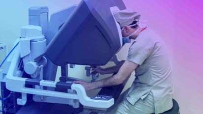
So after the soft tissue component of the flap is inset, we bring in the microscope to anastomose the fibular artery and vein to a recipient vein, artery and vein, in the neck. So the flap can be reperfused. So this is the carotid artery in the patient's right neck. The hypoglossal nerve is at the top of the screen, towards the top of the screen. And the vessels the instruments are holding right now are the vessels from the fibula. Vessels are being dilated prior to the anastomosis.
So once the vessels are prepared, we bring them in apposition. This looks like the facial artery, which is a great artery for revasculariztion in the neck to use. It's being hooked up to the fibular artery here. So these arteries are about three millimeters in size, or just for our perspective.
So once the vessels, the artery and vein, are sutured together, the clamps are released, which allows reperfusion of the fibula. Once we assure that the fibula has been perfused adequately, the neck is closed and the patient will be taken to the intensive care unit.
A typical post-operative course for these patients is they spend three days in the intensive care unit and then approximately three days on the surgical floor. They'll be discharged approximately one week after completion of the operation. We don't allow them to eat anything by mouth until the surgical wounds have healed, which is about a week.
A lot of times these, patients will also have trachs. We'll keep those in place for anywhere between five and seven days, and try and get the trach out prior to patient being discharged. Once the patient is completely healed from this operation and found to be free of any recurrent cancer, the fibular bone can be implanted by an oral surgeon so the patient can have an implant-associated denture for dental restoration down the line. So this is just one of the many complicated head and neck cancer cases and reconstructive cases that we do here at Sentara Norfolk General in Eastern Virginia Medical School.