Chapters
Transcript
[MUSIC PLAYING]
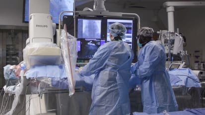
JOHN HUMMEL: This patient is a 62-year-old man who had a prior AFib ablation, and then he had recurrence of his atrial fibrillation that was quite symptomatic, provoking heart failure and hospital admission and was placed on dofetilide. And he had recurrent atrial fibrillation despite the addition of dofetilide.
Given the failure of antiarrhythmic drug therapy and the low likelihood of success on an alternative antiarrhythmic, he was brought back to the electrophysiology laboratory for a repeat ablation.
You can see here that there is a wand-like or multifingered catheter being waved around the heart. This is a multipolar catheter that is collecting electrical signals and electrical voltage from the heart. As we get the electroanatomic map built using the pen array catheter, we see very clearly that the pulmonary veins in red are completely electrically isolated, and there is a residual line of tissue between them that is a different color-- purple, green, and various colors-- that represent the residual electrical activity that was not ablated.
The posterior atrium is largely dead except for a small line of tissue, and so it's very likely that the drivers for atrial fibrillation sit elsewhere. And so you can see us moving the pen array catheter around and outlining the rest of the atrial structure. Here, I'm just outside the-- on the left here, you can see I'm just outside the right pulmonary veins. And you can see there's a large area of purple here with islands of green, indicating there are some regions of fibrosis here.
Those are often drivers for atrial fibrillation. So when we come back later, we'll be looking very carefully at those regions with the specialized software. Here, they tilt the shape of the heart down as I outline the front wall, the anterior wall of the left atrium. And again, you can see some islands of green amongst the purple-- green and red amongst the purple that are other areas of fibrosis that we'll want to look at to see if those areas are anchoring the atrial fibrillation.
So here, right now, we are outlining where the esophagus sits. The esophagus is the structure we want to try to avoid. If we are ablating with energy near the esophagus, we want to limit the energy. And the device being used here is an electroanatomic mapping tool that's placed inside the esophageal temperature probe.
When we finish that ESOPHASTAR map, we'll go back to mapping the atrial fibrillation. If we do find important sites overlying the esophagus, we'll lower our energies and carefully measure temperature to make sure that we don't end up with a risk of esophageal atrial fistula.
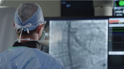
And you can see here now we have almost a complete outline of the heart. And we are looking at various sites. And we will start collecting data. You can see the underlying atrial fibrillation on the right screen there. And so I am looking here for regions where we can get adequate electrical recordings to use the CARTOFINDER software to identify drivers for atrial fibrillation.
And most of the areas where I'm moving the catheter at this point are clearly-- have no electrical activity. If you look at that screen on the right, the two red lines, below the red lines are the recordings from the PentAray, from which we'll do CARTOFINDER mapping. And you can see there are almost no electrograms on those multiple lines-- on the 20 poles from the PentAray-- meaning that all the areas I've looked at so far appear to be electrically inert, at least from the endocardial standpoint.
Here you see we're in atrial fibrillation, and there is a green dot in the RAO projection on the right. It shows an area right in the carina between the right superior and inferior pulmonary veins that the CARTOFINDER programme identified it as potentially important. So we move our ablation catheter into this area. And in doing so, you can see the rhythm on the right will convert to sinus rhythm, just through bump ablation of the region.
Clearly, this appears to be a potentially important site for ablation. So we came back and ablated this region to eliminate that region of fibrosis and reentry that appears to be helping sustain AFib in the left atrium.
Now, one can appreciate the map on the left here. I've already ablated the posterior wall, because the posterior wall, even if you don't identify it on CARTOFINDER or with other mapping systems as being important for sustaining AFib, we know from prior surgical literature in persistent patients that it is an important substrate for developing atrial fibrillation, either now or subsequently. So we made the decision to homogenize what was left in that small region of the posterior wall.
On the right here, we're just eliminating this whole region. So we've done the posterior wall. We've done what appears-- we've ablated what appears to be a critical region in the carina. And at this point, we will reinduce atrial fibrillation if possible, and then we'll probably move on to the right atrium and see if we can map any significant drivers in the right atrium.
The work that has been done on extrapulmonary vein drivers, there are a couple of manuscripts that suggest that patients with obstructive sleep apnea have a higher incidence of extrapulmonary vein drivers from the right atrium, which makes sense because they tend to have higher pulmonary pressures and subsequently larger right atria. And this patient, in fact, does have significant sleep apnea.
On the right side of the left screen, you can see the lesions that were placed around the CARTOFINDER area that allowed restoration of sinus rhythm. And you can also see the posterior lesions that were placed to eliminate the residual voltage on the posterior wall.
And here we get a good view of the posterior wall. Again, looking at the left handed screen, you can see on the left side of the left handed screen the posterior wall lesions, and on the right screen the CARTOFINDER regions. And we continued to look for more sites in the left atrium after induction of AFib but didn't find any and decided it was time to move on to the right atrium.
Now we're finished with the left atrium, we've induced atrial flutter, and we're going to map out the right atrium. And here we're creating the shell for the right atrium. Here we come over to the region of the mitral valve isthmus, and we'll pace here to make sure that there is no evidence that a flutter is being driven by the mitral valve annulus.
And we did entrainment in this region. Found that not to be the case. We did find another driver in the left atrial appendage. But given the increased risk of potential thromboembolic events with ablation of the left atrial appendage, we elected not to pursue that and decided to withdraw, go to the right atrium, and see if we could find drivers in that region.
Now we have this very organized atrial fibrillation. It's no longer atrial flutter. It's variable in cycle length, but it's quite organized. We don't find any drivers in the left atrium for this. So we are now creating the structure of the electroanatomic map of the right atrium. And you can see us build out this map.
It's all been built out. And here you see some flashing going on. That is a replay of the CARTOFINDER map. You can see that there is an area that flashes a bright white intermittently. And that flashing-- that area that's sort of a bright white appears to be a region of focal impulse that may be driving atrial fibrillation.
So in this next screenshot, here you see us positioning the ablation catheter to try to do an ablation over this region of focal impulse. After ablating that septal portion, we are able to reduce atrial fibrillation, although it's fairly organized, and decided that it would be best to target this region of the superior vena cava just opposite to the region that we identified at the base of the right atrial appendage.
Again, we wanted to avoid ablation inside the appendage. Decided to go after the superior vena cava region first. So after mapping the phrenic nerve and making sure we wouldn't insult phrenic nerve conduction, we ablated in this region of the superior vena cava, which restored sinus rhythm again. At that point, we were unable to induce atrial fibrillation again at baseline or [INAUDIBLE], and we called an end to the procedure.
The patient is now two months out from his ablation without atrial fibrillation. Has come off antiarrhythmic drug. But given the degree of scar that we see in the atrium, we are doubtful he'll be able to maintain sinus rhythm without antiarrhythmic drug on board. So we'll see. But it's worth trying to get rid of antiarrhythmic drug therapy if we don't absolutely need it.
My suspicion is that he has enough substrate that he'll develop atrial fibrillation again and that with cardioversion and reinstitution of antiarrythmic drug, that the work we did hopefully will maintain sinus rhythm going forward.
John Hummel, MD, performs a complex ablation on a patient with persistent atrial fibrillation.
Related Presenters

Clinical Cardiac Electrophysiology and Cardiovascular Disease
Professor of Cardiovascular Medicine
Related Videos