Chapters
Transcript
The procedure we're talking about is VT ablation or ablation of ventricular tachycardia.
And the clinical need is really, I think, underserved in many parts of the country.
Emerging data from multiple centers, including centers I've been a part of and our own centers with evolving data, is that patients with recurrent ventricular arrhythmias do worse.
They have higher mortality.
They have more heart failure admissions and hospitalizations.
And so can we alter that morbidity and mortality curve by intervening sooner to address ventricular tachycardia?
Most commonly, the only definitive therapy for VT is a defibrillatory shock device implanted in the chest that can deliver life-saving shock in many instances.
In some cases, it can pace the arrhythmia to terminate it.
But either way, outcomes are worse when those arrhythmias are present.
And so the procedure we do is meant to reduce the burden of that arrhythmia.
In this part of the country, for the most part, VT ablation has not been so commonly done.
And the general practice has been to use medications and to continue to escalate doses of medications to treat the arrhythmia until they fail.
And it turns out that that approach is associated with oftentimes worse outcomes from a heart failure perspective and higher mortality, at least in the ischemic population, the ischemic VT population.
So our procedure is complicated.
It takes several hours, and there are risks.
But we do a good number of them.
We're the highest-volume centers now, certainly in the southern United States, and among the highest volume in the US with what we're doing.
VT ablation is indicated in patients of two varieties.
And when we think about ventricular arrhythmias-- and I actually just wrote a chapter for the American College of Cardiology board review course on VT management.
And what we generally teach is that we first discriminate between those patients who have ventricular arrhythmias but normal hearts.
And those folks generally tend to have good outcomes.
And VT ablation can be curative in that population.
And then the other group of patients are those that have structural heart disease or cardiomyopathy, failing hearts.
And in that population of patients, ventricular tachycardia can portend a worse prognosis, a higher risk of sudden death, a higher risk of heart failure death.
In either case, there's a benefit to the procedure.
On the one hand, it's curative.
It makes people perhaps better, with either no need for medication or a much reduced need for medication.
And in the sicker population, those with structural heart disease, reduces the pain and suffering associated with defibrillatory shocks, reduces heart failure admissions, and we think improves mortality.
What we do, it's not unlike, for those who have heart disease who've undergone cardiac catheterization, where an interventional cardiologist will place tubes up to the heart and shoot dye, squirt dye, into the coronary arteries to create a roadmap of those coronary arteries, from the perspective of the patient, it's not much different.
We still get access from the groin, from the arteries and veins in the groin, which provide a conduit, if you will, up to the heart.
And then when we get up to the heart, we're able to put our catheters into the chambers of interest, either the left ventricle or the right ventricle.
And with the catheters in place, we create very dense, very rich, detailed, three-dimensional anatomic maps that outline the presence or absence of diseased areas of the heart or scarring of the heart.
And scarred heart muscle tissue has what we find to be short circuits that create these fast heartbeats.
And so through a variety of signal processing methods and with our mapping tools, which we've helped to develop, we are able to begin to not just see scar, but we're able to actually visualize the physiology of that scar.
What parts of that scar are problematic?
And what parts of the scar are things we can ignore?
And not so long ago, we couldn't distinguish between the two.
We just saw scar.
And so we're getting smarter in how we map these areas of interest.
And what it means is that we can perhaps be more selective in how we ablate.
It's less procedure time, and with that, we think better outcomes.
What we're doing largely involves very complex interplay between nursing staff, technical staff, oftentimes our heart failure group, because many of these patients have advanced forms of heart disease and are either under evaluation for left ventricular assist devices or transplantation.
And so we co-manage the patients with them.
And then just the duration of the cases is non-trivial.
And in many instances, some of the electrophysiology laboratories are expensive places.
They're risky places.
And you need a lot of experienced people to pull together all the resources that are necessary to deliver not just safe care, but optimal outcomes.
Our team is pretty well versed.
We've worked very hard over the past two years since I've been here to build the team.
It's really good now.
And it was good a year ago, but it's way better than it was a year ago even.
And we continue to try to improve.
And part of our academic credo is that we're always trying to understand how best to do things or constantly reevaluating ourselves and our practices.
And we participate in studies to best understand how to use and develop some of these tools.
And we are fortunate.
We are the first center in South Carolina, we are the first center in the entire southern United States, and we're the second center in the United States to perform this procedure with this mapping technology.
And we're proud of that.
And that is something that we've worked hard to be recognized for and to be given those opportunities.
And because of this kind of work that we do, we are invited to participate in research studies.
And we're in the process of getting a lot of our own experience out in the form of publication with some of the mapping workflows that we've developed here.
The really revolutionary part of this new mapping technology with this grid catheter is that traditionally in the past, we would use two electrodes to create maps of these ventricular chambers and to discriminate scar and to look for some of the interesting signals, what we call late potentials or LAVAs, which stand for local abnormal ventricular activities-- LAVAs and late potentials.
And we didn't do such a good job identifying them or discriminating them in scar with just a point-by-point workflow.
And one of the problems, among many, but one of the problems was that the ability to see scar was dependent upon how electricity flowed through the tissue, what the wave front.
If you can imagine an ocean wave hitting a catheter like this, sitting like this, it's going to hit it kind of with full force.
If it's sitting like this, it's going to blow right over it.
And you're going to see less of the force.
You're going to see less signal, if you will, depending upon how you are oriented to that wave front.
With this catheter, with this grid catheter now, not only are there more than two electrodes, it's a four-by-four arrangement of electrodes that are very small.
So it sees only a very small local environment, rather than sort of a larger body of water, if you will.
It sees a small environment.
And it can look in both directions now.
So the wave front orientation is less relevant to what we see.
MUSC has done a very nice job staying ahead of the curve.
When I have gone to some of the people in leadership, saying we need to be able to do these things, we need to be able to offer these catheters.
And they're not being offered to everybody.
And it means we assume some risk in adopting this technology earlier.
Now granted, we've done a lot of work behind the scenes.
We've done a lot of research work, not in human patients but in animal models.
So we've done our due diligence.
We've done our homework.
But then when it comes time to really testing these ideas in humans immediately after the FDA approves the technology, there's still not a lot of experience with it.
And so we are putting ourselves in a position to really lead the discussion about how we utilize this technology.
This is why we're here.
We're not here to do things that are done every single day.
We do things that are done every single day all the time and with high quality, high reliability.
But we're also here to push the field forward, to innovate, and to make patient care and safety better for everyone, not just in South Carolina but through the southern US and across the US.
And that's the reputation we hope to have.
It's the reputation that's evolving.
We're proud of that.
And the hospital is putting us in a position to be able to do that.
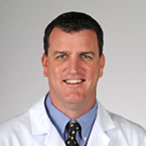
Jeffrey Winterfield, M.D., Hank and Laurel Greer Endowed Chair in Cardiac Electrophysiology at MUSC Health, discusses the new HD Grid procedure and how it impacts patients and allows surgeons a more detailed view of the heart during surgery.
Related Presenters

Jeffrey Winterfield, M.D., is an Associate Professor of Cardiac Electrophysiology at the Medical University of South Carolina. He received his undergraduate degree from Amherst College in Massachusetts and his medical degree with Honors ...