ZACHARIAS P. TSIAMOULOS: Thank you, Professor Gustaudt, our dear chairman. Thank you very much for the kind invitation. I just need to really express my deep appreciation to Stavros and his whole team to invite me here to give a very brief talk about this innovation in colonic dissection. It's for me, a great honor, because I elaborate some of the information about [INAUDIBLE] of my PhD at Imperial College. So I was really blessed to see that from the bench to the bed, so as it translates from the study.
So I'll get you through very quickly on slides I'm sure you've seen before. So this is the first time that there was a diagram presented in Nature Reviews by Prof. Saunders and myself, where we managed to delineate where are the margins for EMR and for ESD.
And that was followed by a study that was published in endoscopy a year after where we proved that when a patient-- when the resection defect was above the vessels, there is sudden burst of vessels that predict a low risk of delayed bleeding. And as we know, the colorectal ESD achieves a very high en bloc resection with a low recurrence rate. We can have accurate histology. But-- there's always a but. And it's a long procedure, time-consuming, and it harbors a high complication rate.
There is an evolution of colorectal ESD even in Japan. And you can see this is the same group that, after 10 years, you can see that they have definitely improved their time, the en bloc resection from 88 to 96.9. They reduced the time of operation, and with a similar complication rate.
So if the Japanese actually improve, we have a long way to go in the West. So I think, for colorectal ESD, what I found for the last three years of my life, you need to be dedicated, committed, and to have a team surrounded by you, by the surgeons and by nurses. And we have to be able to identify-- to have the ability and training to identify the type 2B with zoom scopes, which then, with the interpretation of the pit pattern, to move to the colorectal ESD section and remove a high-grade dysplasia or sm1 lesion.
And my colleague previously was mentioning by the AI, Artificial Intelligence, now we are-- this is the endobrain from Professor Kudo and Dr. Mori. It's a courtesy slide. I was there last year. And that's why we have to understand and actually assess the vessels using the narrow-band imaging. And also at the same time, we have to be able to assess the nuclei and also interpret any atypia.
And so this is the endobrain. And now this is going to move forward to the large polyps. And we have to be also familiar with the pocket creation. It took me a while to understand why my professor, Professor Saito, in the National Cancer Center, was doing that under its reflection, because it gives you a better platform.
And we have to actually be able to understand and comprehend what a pocket means compared to the conventional and the physical standard flap. We have to be in the position of using all the accessories when it is necessary, when the fibrosis nylon clip on the left colonic lesion, SO clip again on the right colonic lesions.
And tunneling, which is not very commonly widely used in the colorectum, because there are not so many publications. But it's definitely a forward for the point procedures. But I think there is a trend now to implement tunneling in the colorectum.
So in UK-- that's why I kept still the symbol of the EU for two more months-- actually, we don't have all this ability. We have lack of optical imaging. We have lack of training, because you need to be dedicated. So we'll have these complex polyps clinical governance meetings. And we discuss the case with surgeons, pathologists, and radiologists. And then some of these cases, they've been streamlined for colorectal ESD.
So these are the four main issues, I think, that every Western operator has always in his mind about the safety profile, about the time of the procedure, about which technique approach, and the learning curve. So I'm very, very happy and very honored to present the first Western bipolar device. But before we go in this direction, I just want to refresh your memory. Currently, we're using monopolar radio frequency energy for cut and coagulate small vessels.
And we're using bipolar devices to coagulate only for coagulation of large vessels, because with the monopolar, there's a potential of remote burns. And with the bipolar, there's a minimal risk of remote burns. And these other devices-- as you know, everybody is familiar with these devices. But Professor Hancock and his team, in 2009, they wanted just to think outside the box.
So I just want to refresh your memory. When we use a knife, a snare, any electrocautery device, the range is between 200 and 2000 voltage, because it has to go through the tissue impedance. And that's where you need a lot of energy.
So the team from Bangor thought about the advanced energy. So they had to think outside the box. So they went below the 200. And they created the microwave for calculation and a cut for bipolar. So this is the microwave energy, which is in a way, it is an irradiating electromagnetic field, which, in a way, it oscillates the ions within the molecule. And that can cause vibration. The vibration can cause thermal heating conducted to each cell. And that's why we have this tissue effect.
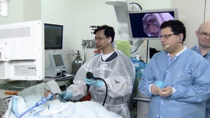
And this is really related to the design on the instrument, to the time of the application, and to the high frequency. High is the frequency. Then lower is the depth of the penetration. So we have the "Speedboat," which can inject. It can cut very precisely between the two upper blades. Stavros is going to use the Speedboat later on in one case.
It can coagulate vessels with the radiation of the microwave modality, again, very clean coagulation. It can fully rotate. This is deja vu, Professor Gustaudt, I'm feeling. Four years ago, I was at the [INAUDIBLE] in London when I was presenting the Speedboat. And there was a question of yours about why I was actually pointing the tip of the device, because at that time, we didn't have the rotation. And it took us about-- I don't know for how long-- four or five years to achieve that.
And so this device now, we have done successfully a preclinical GLP study published in Endoscopy. We proved that there's a viability of the muscle. And we had to go through a proper human trial to prove the safety and efficacy of a microwave on the precoagulation and coagulation of the vessels.
And this the first publication, which is coming soon in the next two weeks, where we removed a polyp in 25 minutes, of 4 centimeters, using the ZoomScope to assess the type A pit pattern, a G-net, and then using the Speedboat with short bursts, we prefer the tunneling and the coagulation of vessels. You can see now, this is a big feeding vessel, which is we have to coagulate up to 10 seconds, because then every second, you augment the energy by 10 joules. You see this bubbling, blanching. And then it's very efficient coagulation.
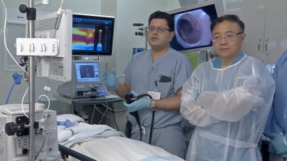
And then we move to the tunneling. You can see how precise. After contact, you deliver the energy. And that polyp was successfully removed in 25 minutes with no uneventful complications. You can see, we had the in and out approach, which is opposite to the IT NanoKnife, because it has similar dimensions with IT NanoKnife.
And now the bleeding, I'll just leave that to play now. You can see, with tamponade, with the optimization of the angulation of the device. And you leave that on the vessel for up to 10 seconds. And this is very, very successful. Sometimes you have to go a bit the second time and third time. But that's the essence of this.
And that's the last bit. I just want to go into the last bit and show you another bleeding, which was successfully stopped. And this is the last bit. We can see, the defect has no carbonization, no charring. And you just see a clean and precise cut.
So you can see how the submucosal has been sustained and viable without any damage to the submucosal layer. So the features of the Speedboat, it has to be in contact and can really deliver energy in a confined manner. It should be used in an in and out approach, always parallel to the muscle layer, and apply microwave more than 6 seconds, up to 10 seconds.
So the first case, it will be published soon, have 24 lesions in three weeks. And we had most the LST G type. We have three T1 cancers. But two of them had endoscope surveillance with no recurrence. The third one was an old lady with more than 1,000 microns. But because of the high of comorbidities, she had to decide to follow that.
And we did under sedation, mainly. You can see the hours. Stavros has his own algorithm. But I think this is very important. We have half of the cases that are done under half an hour and under two hours under sedation.
And this is a sign which is in one of the cases which I did. I had to abandon because of the muscle retraction sign. So I had 67% of en bloc dissections. And I had to convert, in some cases, on a Snare, because in NHS, we cannot have spent nine hours doing ESD.
And we had no complications, maybe just one or two cases of delayed bleeding, no perforations, no microperforations. Microwave was successful in 89% of the cases. 80% of the patients, they were sent home the same day. And from them very small case series, we can say that lesions less than 5 centimeters, that we can achieve en bloc dissections in less than two hours by using the tunneling approach. Early days, but it looks very promising.
So this device might be able to address some of the previous issues about design, the novelty ESD in less than two hours, possibly using tunneling or pocket and a dedicated training and mentorship program.
Thank you very much for your time. And I just want to also invite you in our course that we, last year, were the first that we had the live cases with a Speedboat. And also, we had the honor to invite Professor Saito, who used also the IT NanoKnife. Thank you very much for your time.
[APPLAUSE]