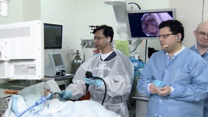
HARUHIRO INOUE: Thank you very much, Dr. [INAUDIBLE], for your kind invitation to this great course. Today I would like focusing on to the GERD after POEM. So world's first case of POEM was done in 10 years ago-- 11 years ago.
So this actually is a case-- this was actually the case and the before treatment. And then they place anterior myotomy like this. And then I perform the circular myotomy. Then, after completion of the circular myotomy, we close our mucosal entry clips.
So this is a photo of the third case. The first case we didn't have. Anyway, so this is before-- the first case before and after procedure. So volume pass [INAUDIBLE] symptom score improved very well.
This is the same patient, first patient. We met together eight years after POEM procedure. He said 100% satisfaction. And he can we eat well and gain weight, 20 kg. And no GERD symptom. So this is an endoscopic image a eight years after procedure. So his junction is open and no reflux.
So far in our hospital, we've performed 1,900 cases. And fortunately, our success rate is 95% and more. And there are no major complications. So we are very lucky.
But some of the patients has a GERD symptom. So GERD symptom itself, 20%. The erosive GERD is 25%. And the abnormal pH is 50%.
So we have to think of something to control the post-POEM GERD. So first is we are thinking is rings of gastric side myotomy. It's like this. So once we place more than 4 centimeter myotomy, even it's anterior or posterior, so we may, might cut the whole collar-sling muscle. Then patient becomes the severe GERD. If we place a more than four centimeter myotomy in the [INAUDIBLE] then so some patients becomes a highly risk of grade C esophagitis.
So simply, we have to avoid too much cut to the gastric side. And then control the rings of the gastric myotomy. Double scope technique is our most available useful technique. Some Portuguese doctors reported the double scope technique.
We accepted this procedure in the old cases. But today, in the lab demonstration, we can demonstrate. So we control the-- we can recognize, using the intragastric scope view, we can see submucosal endoscopy tip light.
Then we can measure the gastric side myotomy rings exactly. So one to two centimeter or scope diameter the best. And then if we dissect three centimeter, four centimeter, high risk of becoming GERD.
And another technical point is the preservation of the collar-sling muscle. So everybody knows gastric his angle is made of collar-sling muscle, existence of a collar-sling muscle. So, we are trying to not to cut the collar-sling muscle. Of course, collar sling muscle, there is no collar-sling in the [INAUDIBLE]. So we control the direction of gastric myotomy. Posterior myotomy, without [INAUDIBLE] oblique muscle, 66% patients becomes GERD. But if we preserve collar-sling muscle, the risk of GERD reduces.
Yes, once we cut the oblique muscle, the risk factor for GERD C esophagitis becomes higher. This is actual image. In this case, we approach posterior esophagus.
We can see the edge of the collar-sling muscle as a yellow dot. And then this the [INAUDIBLE] muscle. And then in between, we can identify perforating arteries. Then though we will go to the little bit right-hand side.
On the left hand side is an image of a patient from an ESD, cardiac ESD, not a POEM. But anyway, we can recognize the no [INAUDIBLE] curve. There is no collar-sling muscle. So we control the gastric side myotomy direction to the [INAUDIBLE] curve of the stomach like this, OK.
So in a double scope technique, if we recognize the light from the submuscosal endoscopy if it's located at the [INAUDIBLE] curve, that means we succeeded to preserve the [INAUDIBLE] muscle-- collar-sling muscle.
OK, so like that. So that is the very simple technique to preserve the collar-sling muscle. So another one, another one is the endoscope [INAUDIBLE] during a POEM procedure we perform the anti-reflux fundoplication. That is a POEM plus fundoplication.
So technique is like this. We approach the GE junction anteriorly. And then after creating anterior tunnel beyond the diaphragm, we get in abdominal cavity directly. Then we make a partial anterior fundoplication.
Actual procedure is like this. So for us 40 patients received this procedure. So this is the end of myotomy. Ah, so like this, if we perform a layer POEM procedure, before procedure, junction was tight.
But after procedure, junction open. Then the patient becomes-- potentially becomes a reflux disease. Then this is the submucosal tunnel in the anterior wall. And the to myotomy has already been dammed. Then at the end of the submucosal tunnel, below the diaphragm, we are trying to get in abdominal cavity. So behind, we can see back side of the left liver lobe.
We dissect the peritoneum and then get in their peritoneal space. On top is an abdominal wall and the bottom is a anterior stomach, pharynx. Then one of the technique is a catch, the anterior gastric wall using loop and crimp technique.
Place three or four anchors, distal anchors, to the anterior stomach. And then after that, we also place-- some proximal anchor to the distal end of the submucosal tunnel. Then this is a placement of the second the clip at the distal end of the submucosal tunnel.
And then if we close the end loop, we can complete the partial fundoplication. So this is actual image. Now we are trying to close the end loop. And then right-hand side, you can see fundoplication is creating now. OK, so left-hand side is before fundoplication, endoscopic fundoplication.
So right-hand side is after endoscopic fundoplication. So this image is very similar to a dull fundoplication. OK, so, of course, we can perform this procedure using a suturing device. So we catch the anterior wall of the stomach, gastric [INAUDIBLE].
OK, now I'm [INAUDIBLE] the left live lobe [INAUDIBLE] away. OK, so I need these out. And then so first distal stitch. And then after that, proximal distal end of the esophagus, the second stitch. OK, so one more stitch.
So we use a berb snare. So just suture and the pull it, it's enough. It's not necessary to tie over. Then we can make like this fundoplication in the submucosal tunnel. So this is right after procedure. Patient phonics looks more tight than just the POEM.
So this is data. At the time, over 31 cases. Procedure was successfully completed in 100% cases. And there's no adverse event. And the ILP and the Eckardt score improved.
You don't see esophagitis is controlled better than POEM alone group. And the other [INAUDIBLE] study looks getting better, but no statistically difference. We need more cases.
So ladies and gentlemen, this is my conclusion. So often we have achalasia patient, previous endoscopy checkup, the patient has no hiatal hernia. We perform a posterior POEM. And then the patient becomes GERD.
So at that time, we perform very few case, I think. So we perform the POEM F, per-oral endoscopic fundoplication in the anterior wall of the esophagus. And the patient who has-- achalasia patient who has a high rate of hernia, we directly perform the anterior myotomy plus fundoplication. That is our strategy. Thank you very much for your kind attention.
[APPLAUSE]