Chapters
Transcript
For this particular case, this gentleman had a pituitary tumor-- a very sizable pituitary tumor-- that had been really just partially resected by an outside surgeon, and he still was very symptomatic.
General symptoms include a headache or malaise or sinus complaints or what have you.
Then there's very specific things that relate to the hormonal function and compression of the tumor itself.
So pituitary tumors can be non-functional or functional.
Non-functional tumors, if you have a sizable tumor, can compress the normal pituitary gland and cause it not to work like it's supposed to, so you can wind up with low levels of pituitary hormones in general.
Then, if you have a functional pituitary tumor that makes excessive hormone, that can also present clinical symptoms depending on what the hormone is.
Then you have compressive issues, meaning from the size of the tumor.
So the optic apparatus runs very close to the pituitary gland.
A very sizable pituitary tumor can give you vision loss from compression.
Characteristically, it gives you peripheral vision loss first, but it can-- I've seen it favor one eye over the other and a variety of different vision loss patterns.
But that can be one of the symptoms we see as well.
This tumor was a non-functional pituitary tumor, so not able to be treated with medicine.
Really, surgery is the main treatment modality.
And he needed another resection.
So we offered him a resection.
The way that we usually do that is with an endonasal resection.
So instead of opening the skull and going in from the side or the front of the cranial cavity, you go through the nose to take these tumors out.
That operation is a combined operation.
We do that surgery with two teams of experts.
So we do the operation with the ENT surgeons, the rhinologists in particular.
So they do the nose portion for me.
So they clear a passageway from inside the nasal cavity back to the skull base.
And that's stuff that they do every day.
They do it with endoscopes, and they're operating on patients with sinus disease and so forth and sinus cancers.
So they clear the passage back to the skull base, and then we work as a team to remove the tumor.
And again, we do it with endoscopes, so it's a [INAUDIBLE] camera on the end of a small tube.
Do the operation through the nasal holes, and we watch the computer screen as we do the operation.
We have small dissecting instruments to remove the tumor from the delicate structures around it-- both the normal pituitary gland as well as the optic nerves and the carotid artery.
Sometimes with sizable tumors, even the base of the brain.
That's a tedious process, and once you're done, you have to fix that hole.
So we have a multi-layered way that we repair that hole in the skull base, both with material that is artificial-- so allograft-- and also some tissue from the patient's nasal cavity.
We use part of the nasoseptal mucosa and turn that as a pedicle flap to cover the skull base as a vascularized flap, so a nasoseptal flap.
So the ENT surgeon initially clears a passageway through the nasal cavity.
So they often raise nasoseptal flap first, especially when we're quite sure that we're going to need it.
They clear the anterior aspect of the sphenoid sinus and make a sphenoidotomy open into the sphenoid sinus and they clear the sphenoid septations.
And then we work together at that point, once we get past the opening of the sphenoid sinus.
So we work through both nose holes-- both nares-- with, again, small bayoneted instruments, and do the operation as we watch the computer screen using the endoscopes.
We remove the bone of the anterior face of the sellae, widen that as wide as possible to be able to have a clear view of what's going on.
The idea of keyhole surgery is that you have a small opening and you're able to get nice and close and see very widely on the other side, but you want to be able to see widely, so you try and make that hole as big as you can.
So when we look through the small aperture of the nose, you still need to see widely inside to see what's going on.
So remove the face of the sphenoid sinus, face of the sellae, open the dura over the tumor.
And again, in this particular case, we had to deal with a fair amount of scar tissue that was there from the prior surgery.
And then you use small curating instruments-- suction devices, small scissors-- to remove the tumor in piecemeal fashion.
Sometimes with smaller tumors, you're able to get around it and completely remove it on block.
With very large tumors, we often have to do that in piecemeal fashion.
So as the tumor-- we compartmentalize our resections.
We work on the bottom and the sides and the top, and then try and remove the tumor from the diaphragma sellae, which is the division between the pituitary gland and the brain space, the suprasellar space, out of the cavernous sinuses on both sides, and to the posterior aspect of the sellae posteriorly.
And once we're able to see all those areas, we turn our attention to the reconstruction.
So we use, again, artificial material to seal over the face of the sellae.
We often use a cloth-like material, and then often a resorbable plate to give some rigid reconstruction.
And then we use some of the mucosa from the inside of the nasal cavity off the septum as a nasoseptal flap as a vascularized repair.
Then, the ENT surgeons often pack the nose with some packing at the end to be able to hold everything in place.
So patients often stay in the hospital three-ish days after the operation.
They do so for several reasons.
You have to make sure that all of the hormone levels are what they're supposed to be, so there's lab work that's involved with that.
And it can take a few days sometimes to sort all that out, whether it's getting the right dosages for medicines that need to replace, or making sure that the pituitary is working like it's supposed to.
We often have packing that's in the nose, also, from the closure from the ENT surgeons, and that often stays in a few days and needs to come out.
We like to do that in hospital if we can.
Then, in addition, we need to make sure that there's no spinal fluid leakage.
So one of the reasons for a vigorous and robust repair of the skull base is to prevent spinal fluid leakage.
And staying in the hospital till the packing comes out and then making sure there's no spinal fluid leakage is important because that's a very serious thing if it happens.
The patient did very well after surgery.
He just stayed a few days in the hospital, and I've seen him back in clinic since then and he's doing quite well.
He unfortunately had a stable vision loss, meaning he had significant vision loss prior to surgery-- been there for quite some time before he came to see us, unfortunately.
So he did not have a dramatic improvement afterwards-- maybe a small improvement, but not a dramatic improvement.
It's always good to see patients sooner if we can, even at the first surgery, so that we can try and get all the tumor out in the first operation if possible.
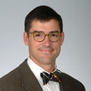
Alexander Vandergrift, M.D., a neurosurgeon at MUSC, describes removing a nonfunctioning pituitary tumor from a patient through endonasal resection using an endoscope.
Related Presenters

William Alexander Vandergrift, M.D.
William Vandergrift, M.D., is an associate professor in the Department of Neurosurgery at MUSC. He trained at the Medical University of South Carolina for neurosurgical residency and at the John Wayne Cancer Institute in Santa Monica, ...