Chapters
Transcript
SATISH NADIG: My name is Satish Nadig. I'm an Associate Professor of Surgery in the division of Transplant Pediatrics Critical Care and Microbiology and Immunology. Initially, during the beginning of a laparoscopic donor nephrectomy, we make a six to seven centimeter incision around the umbilicus, depending on the size of your hand. And a port is placed in the left upper quadrants and left lower quadrants, both one centimeter or 10 millimeter ports. Here, we take down the ligamentous attachments from the left colon to the abdominal wall up towards the spleen using a Thunderbeat energy device, although any energy device can be used.
Once these attachments are released, the left colon is then taken off of the abdominal wall by following the peritoneal reflection of the white line of Toldt. This is carefully dissected such that the retroperitoneal fat is separated from the mesenteric fat along this white line of Toldt, which you can see the instrument carefully follows.
Once that is completed, the dissection is carried down towards the pelvis with both blunt and sharp dissection using the energy device for hemostasis. We do this until we feel the pulsation of the iliac artery as a stopping point. We then identify the ureter and encircle that safely with our finger by protecting it, pulling it medially, and dissecting the tissue laterally. Often, there will be a gonadal accessory artery, which you can see here, that can be taken with the energy device.
We then stay in the safe plane lateral to the ureter and follow this plane up to the lower pole of the kidney. You can see here that we've identified the gonadal vein and are staying lateral to these structures until we are able to identify the lower portion of the kidney itself. Once we have taken the dissection up to the lower pole of the kidney, we separate the ureter from the gonadal vein.
And here you can see the lower pole of the kidney clearly under my finger. We then use the gonadal vein as a landmark once the ureter is separated off and stay anterior to the gonadal vein by taking branches of it sequentially up to the junction of the renal vein. This can all be taken with the energy device, which maintains hemostasis throughout the procedure.
Care is taken to see if there are any lumbar veins that are coming from below the gonadal vein from the renal vein itself. Also, it is in this area below the gonadal vein that a lower polar artery often runs if one is identified. In this particular patient, a lower polar artery was not seen on preoperative CT scanning, but sometimes these lower pole arteries can surprise you. So it is important that care is taken when the dissection of the gonadal vein is commenced up to the renal vein.
Once the vascular dissection begins, depending on the energy device that is used, it is important to cool the blade of the energy device prior to resting it on the vascular structure itself so as not to injure the vascular structure. The tissue is clasped and pulled away from the vessels prior to initiating the heat. Once the renal vein is identified with the gonadal vein draining it, the adrenal vein is then dissected free. The adrenal vein is followed high enough such that it can be sequentially clipped and divided, all the while maintaining that the blade is cooled prior to commencing the dissection of the vascular structure.
Here we began to see the beginning of the adrenal vein, which is important to dissect free on both sides such that a clip is securely able to be placed both proximally and distally on the vein itself. The clip is placed and then positioned with space in between enough to transect the vein. Two clips are placed for safety purposes. And the adrenal vein is then transected sharply.
It is here where the artery is often seen as well as the adrenal gland in the superior aspect of the picture. Tissue around the renal vein is freed, and the adrenal gland is separated from the upper pole of the kidney. This dissection is taken down to the psoas muscle, such that the upper pole of the kidney is mobile.
Manual traction can be achieved to expose the tissue between the upper pole of the kidney and the spleen. Care must be taken to not aggressively push the kidney away from the spleen in order to prevent injury to the splenic structure. The gonadal vein is then sequentially clipped and divided.
Typically, two clips usually suffice. In this case, the gonadal vein was quite large, and clips were placed to ensure that the entire gonadal vein was adequately occluded. The gonadal vein is then sharply transected.
Once the gonadal vein is free, it is used as a handle to expose any lumbar veins that are coming from the bottom of the renal vein on the left side. And these are often taken with the energy device if they are small. If they are larger lumbar veins, it is important to clip and then divide them.
Once this is completed, the ureter is pulled medially. The tissue between the ureter and the renal vein below the kidney is divided. And the kidney is mobilized from its lateral structures, thereby flipping the kidney medially. Care should be taken here to be sufficiently away from the hilum of the kidney so that the surgeon does not inadvertently injure surrounding structures from the back of the kidney.
Using the hand-assisted method, the surgeon's hand is used to expose the tissues. And the energy device is used for hemostasis and dissection of the retroperitoneal fat and Gerota's fascia behind the kidney. Again care should be taken to not migrate close to the hilum of the kidney from the back side, so as not to injure the artery. The dissection from the back is now met with the initial dissection we did from the front of the kidney to completely mobilize the upper pole.
Once the kidney is completely mobilized, the ureter is lifted cephalad. And the view from below the kidney is utilized to identify the artery situated behind the vein. In this case, the artery, although originating from behind the vein, turns such that it is anterior to the vein, which is somewhat unusual anatomically, close to the hilum. Yet the origin remains in an anatomically normal position, posterior to the renal vein.
The artery is dissected free from the tissues surrounding the retroperitoneal space down to the level of the aorta, such that the junction of the renal artery and the aorta are seen in order to provide the most length possible for implantation. Here, the tissue on top of the aorta is then taken to expose the origin of the renal artery from the aorta itself. It is very important here to make sure the blade of the energy device is cool prior to dissecting on top of the aorta, so as not to injure the aorta.
The origin of the renal artery is seen clearly here coming off of the aorta. Heparin is given to the patient. And in our center, we also do four doses of mannitol throughout the case. Once the heparin is given and after three minutes, the ureter is clipped and divided distally and transected proximally.
The artery is then taken with a vascular load stapler, and this is the time for cross clamp. The vein is then sequentially taken below the level of the adrenal vein such to identify as much length as possible on the renal vein. Once the renal vein is taken, the kidney is mobilized and removed from the patient.
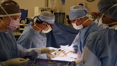
Satish N. Nadig, M.D., Ph.D., surgical director of the pediatric and living donor transplant programs at the Medical University of South Carolina, narrates surgical footage from a laparoscopic donor nephrectomy. In this minimally invasive operation, the kidney is removed through the umbilicus. Because there is less surgical trauma, donors often go home one day after surgery. The recipient of the donated kidney was a pediatric patient.
Related Presenters

Dr. Satish Nadig's clinical interests are centered around adult and pediatric abdominal multi-organ transplantation. This includes liver, kidney, and pancreas transplantation. Additional interests are in laparoscopic donor nephrectomy ...
Related Videos
