[MUSIC PLAYING]
The pineal gland is an organ in the back, middle part of the brain, and sometimes if that gland looks to be more filled with fluid, with distinct borders around it, that can be what's known as the pineal cyst.
The majority of pineal cysts never caused any troubles and they're found what we call incidentally, meaning we're doing the imaging studies to look for other things and we find a cyst on the imaging.
They're very common.
About between 1%-4% of people getting MRIs, we'll find a pineal cyst on an MRI.
When they start becoming a problem is when they are kind of atypical in appearance, and they have compression of surrounding brain structures, and they're larger than typically seen on a incidentally found pineal cyst and they're accompanied with symptoms going on with it.
They can be challenging because sometimes the symptoms are vague, so headache, nausea, vomiting.
It's often rare when it's a cyst to get hydrocephalus, because the cyst component doesn't tend to block off fluid pathway, it just kind of distorts some of the MRI appearances.
And sometimes you can have eye difficulty, eye motion abnormalities.
But often, if those things are present, it could be more than just the pineal cyst in different neoplasms involved.
It's so easy to get MRIs these days.
It's no radiation to kids.
We can even do what we call fast MRIs, where the kids don't have to be put to sleep.
So if you are having a kid with vague neurologic symptoms, it's really easy to get a fast MRI, see if there's anything, not just having a cyst in mind.
And then if the child needs to be sedated for more involved imaging, we can always do that.
Juliana first started, her parents noticed some difficulties when she was about six months of age.
It started where she would be having these head drop episodes, which isn't a typical presentation for a cyst, but she had a lengthy workup for those episodes, including multiple EEGs, long term studies, and those episodes typically go along with a specific seizure pattern.
Her workup for those seizures was negative and it didn't fit the typical diagnosis of that.
The head drops first started in clusters where she would do multiple throughout a day and then they did space out some over time.
But more leading up to when they contacted me was the feeling that she was having a lot of headaches and was in constant pain.
She would stop play and grab her head because she was in discomfort.
A lot of irritability, waking up at night with them not able to console their child, just up for hours screaming and crying, difficulty sleeping, not every night, but increasing in frequency.
So it led them to get additional imaging to find out if there were any answers.
So they had seen providers closer to home and they were told that the cyst couldn't be causing her symptoms and they were very frustrated nothing else they had done was providing any answers.
So they looked online and found MUSC that we have experience in treating pineal cysts and then reached out to see if they could get a second opinion here.
First, I received her MRI studies and review those.
And then after talking with Juliana's parents and hearing her symptoms and reading the extensive workup that she's had and all the negative test results they had to get any answers, I thought everything else has been exhausted.
I think the cyst is likely playing a role here in her condition.
It's hard to tell in kids.
Sometimes pain and irritability is a marker of headaches and that's how she can express herself at that age.
So the irritability and the pain I think comes from some of the compression of the surrounding brain structures.
So these are slow growing, so they are not a neoplasm, they're not a cancer, but they are an abnormal entity.
So it likely over time could have increased in size and caused worsening symptoms.
So it's a challenging location to get to, and of course, you want to operate on the right thing for the right reasons.
Typically these aren't surgically needed to come out, but based on her symptoms and the imaging, I thought it was worth the risk of surgery to offer the chance that it could help improve the symptoms for Juliana.
So we talked a lot about that leading up to surgery.
Even before getting the child to the hospital, just the coordination it entails to go along with it, great nursing staff, getting the patients ready, getting the imaging study, having great radiologists review the pictures.
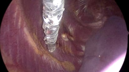
The equipment we use in the operating room is state of the art to do these in a less invasive way to accomplish the goal of getting the cyst out through smaller incisions and less trauma for the patient.
In order to get to these deep structures, you need a lot of magnification and light that helps you get there.
So we have a new technology called the Synaptive Exoscope.
This technology allows us to do it through a smaller incision, do it three dimensionally.
So we use equipment, special glasses, so we're looking at a screen that shows three dimensionally where you're working, and able to do it faster, more efficiently through less incision or a smaller incision for the patient.
You get much better visualization.
You can magnify better, you have better light, the three dimensional component of it.
The whole room can see what you're doing.
So nursing staff, the scrub person can help you anticipate moves.
Anybody observing can see it a lot better.
Ergonomically, from the surgeon, you're in a neutral position and you're looking ahead at the screen to accomplish the surgery.
Whereas, with other microscopes, you'd have to twerk your neck in different positions and it's less comfortable.
So you could do a long operation and feel totally at ease with your body at the end of the case.
So we have special pediatric head holding fixation systems to get the patient in the ideal position.
Make just about maybe a three or centimeter incision.
You do the approach where you go between the two hemispheres and let brain relaxation open up the arachnoid, which is the membranes around the spinal fluid filled spaces of the brain.
You identify the corpus callosum, the tectum, which are both normal things that you want to preserve, and then all the deep draining veins in that area.
You open up the arachnoid around those structures and provide a corridor to get the cyst out.
So you identify the cyst and also mobilize that area and then are able to remove it.
So she did very, very well.
She was in the hospital about three days.
I'd say typical for that type of surgery.
It's anywhere from three to five days that kids stay in the hospital.
She was playful, running around her room, just really doing well.
So she has a great prognosis.
These are benign lesions and her pathology, actually, was a pinealcytoma, not just the pineal cyst.
So that is a benign growth and it's cured if all the mass is removed, it's a surgical cure.
So we will follow her with imaging over the next few years, but by doing the surgery we were able to remove it completely and let her live a normal life.