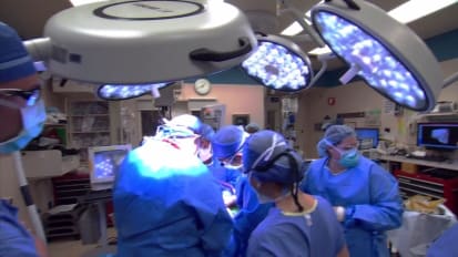
BRIAN KIM: So this is a chest X-ray, and you see broken ribs 5 and 6 on the right hand side as well as a right clavicle fracture. On the three dimensional reconstructions here, you can see the overriding fractures at 5 and 6. The three-dimensional reconstructions are used to plan the incision, and this is the right chest. So this is a right posterior lateral thoracotomy.
Smaller portion of the marked-out incision is made. And the incision can therefore be tailored directionally after the chest wall is defined. See the splitting of muscle on the way down to the chest wall, and then extension of the incision as we have felt the fractures. And this helps, again, central exposure.
So we're feeling over the rib fractures and dissecting down to the chest wall on top of the fifth and sixth rib fractures. We're getting underneath the scapula to elevate it and taking down some of the filmy adhesions. A ring Bookwalter retractor is placed, and you see the angle which helps the scapula to be elevated from the chest wall. It's somewhat subtle on the video, but you can see a bit of fracture hematoma adjacent to the chest wall here.
Now we're opening up over the top of the individual ribs themselves. So the sights of the fractures have been isolated and dissected. Curved joker is being used to reduce the fractures. And a rib tenaculum assists.
Now we're sizing up the fracture and the plate that's being planned to stabilize this fracture. So with the acute RibLoc U Plus System, we need to get into the pleural space over the top of the rib. And you can see that on either side of the fracture, we're entering the pleural space. We're dissecting around the rib, over the top of it.
We're trying to spare any tissue from being on the rib or behind it. Here's the plate with the drill guides in place, and they're being checked and secured to the plate. So the plate in the drill guides are being secured around the fracture. And the drill guides are being secured with this hand-held screwdriver.
And on the side of the drill guide, the depth to screw is measured. We'll have a close idea or approximation of that based on the CT scans as the ribs are measured on the CT. This drill is being used at each of these points. There are four in total. And there's a collar on the drill that prevents it from drilling too far. On either side, we're hand-screwing down the appropriate screws. And then the drill guides are being unscrewed and removed.
We go back and check the tightness of each of the screws. There's a certain appearance to the screws as they're flush with the plate. Often it's just a feel as well. We see that now the sixth rib is being similarly readied for the plate. Tightening down the drill guides. And this will, again, crimp the back of the plate and bring it to the back of the rib, not only securing the plate to the rib but giving us the depth of the screw to come-- drill and screw to come.
Drill guides coming off once again. And final product of five and six. Here's the chest tube being fed posterior apically. It's secured with a O silk suture. We're using EXPAREL as an intercostal field block throughout the visible ribs in the thoracotomy.
A layered closure is performed with running Vicryl. I like to use running number one Vicryl from either side and meeting in the middle on the posterior and then the anterior fascia. In this case, the incision was small enough that a single running layer was performed. In similar fashion here, here's the anterior fascia being closed. Of course, homeostasis is checked for at each layer of the closure.
Here's the post-operative chest X-ray with the plate and screw hardware and the right-sided tube thoracostomy in position. You note that the patient has a dual lumen endotracheal tube in place and the right lung is insufflated, as is the contralateral lung.