Chapters
Transcript
[MUSIC PLAYING]
SPEAKER: So this is really a case I did with metal. It was a case with a-- a pediatric case really who had a CP angle. The CP angle is the cerebellopontine angle. Had a lesion there. It was not really known what kind of lesion that was. The child was in care with pediatric hematology/oncology. And they asked us to remove that lesion.
The only problem was that lesion also caused complete hearing loss and facial paralysis. So what this case is all about is to remove that lesion from the CP angle. So basically, we drill a translabyrinthine approach here. This is what you're seeing. We're drilling the mastoid here. We identified on the posterior aspect of sigmoid sinus. Now we're finding the facial nerve here. You can see the incus lateral.
So we enter the antrum, find the facial nerve. Again, this child had facial paralysis. At the end of the case, we will graft the facial nerve. We do a XII-VII anastomosis. What we can see here, we prepared a sinodural angle. So what I'm pushing back here is the dura. So the linings of the brain are the middle cranial fossa. On the posterior aspect, you have the bluish-looking structure. It's under the suction right now. It's the sigmoid sinus.
We remove all bone from both structures. So for the sigmoid sinus, we push that down. Then medially to that would be to posterior fossa. And superiorly to the left of the picture would be the middle fossa with the linings of the brain.
So we keep dissecting here and find the facial nerve. We extend the facial recess, what's called the facial recess. It's a recess between the actual facial nerve, and the branch of the facial nerve is just called the chorda tympnai, which supplies the anterior 2/3 of the tongue with taste qualities, taste fibers. And so we enter the middle ear here. And we can see here the facial nerve. We put a stimulator probe on it.
To the left of this, we can see the lateral semicircular canal. So part of the inner ear. And so this is all normal anatomy. No lesion expected to be found here obviously.
So we keep drilling here into the labyrinth. We call it a labyrinthectomy. Basically, we remove the inner ear, posterior aspect of the inner ear, and that step provides us access to then what's called the internal auditory canal. So all the way medial here.
So we keep working our way medial. We can see that the lateral semicircular canal here, one of the balance canals, has what we call blue lines. Blue lines mean that we can see the canal underneath the bone. The canal has been opened. We can see the bony opening of the posterior canal as well, right underneath the drill right now.
And we enter the vestibule of the inner ear, which is shown right now. And you follow the superior semicircular canal right here and follow that around. Also push the bone removal all the way posterior into the sinodural angle. And you can see, we can follow that facial nerve, which is right anterior to the suction now. We can follow that all the way inferiorly.
We further remove bone at the posterior fossa here and, again, continue to dissect medial. We usually use a rongeur. This is the smallest rongeur available. It looks giant due to the small size of the ear really and mastoid. Again, this was a pediatric case on top of it. So obviously, instruments look quite large.
Again, continue to remove bone. Again, we pushed the sigmoid sinus all the way back. So right now, we're dissecting on a sigmoid sinus. We can see that wedge of bone is basically in the sinodural angle and really freeing that wedge of bone from all the soft tissues and continue to work medial here.
The important part of removing all the bone here is because that's a craniotomy essentially. So this is what we used to ultimately remove the lesion that we were tasked to do here-- which we were tasked to do. So continue to remove bone. We can see that wedge in the sinodural angle. Again, this all has to be removed.
We are very careful not to cause either a CSF leak or a quite large bleed from the sigmoid sinus. Continue to remove more medially. Now we've skeletonized the internal auditory canal. So this is the area where we can actually then find the lesion that we're here for.
We can see the facial nerve laterally to the right side of the drill. That's called the second genu of the facial nerve, as it runs in its fallopian canal. This is the internal auditory canal. We can see it much better here in a minute.
Again, we skeletonized that, which means basically we take all the bone off of it. And we make it a really soft structure. So 100% of the bone needs to go. Continue this. I skipped forward here a little bit. So we really drill 270 degrees around that internal auditory canal to remove more bone. It's quite a tedious process.
We can see here on the right side of the drill is the jugular bulb. You can see that nicely. We open the cochlear aqueduct, which is viewed right here. And the cochlear aqueduct is a connection that usually contains cerebrospinal fluid. So it's part of the subarachnoid space.
You can see here how we can open this, and we release cerebrospinal fluid. All right. So this is a really nice way to decompress everything, give us some more room. So what that basically allows us is to dissect in a larger space.
So this is the lesion. It's a malignant lesion, which is very rare in this area. So what we did is we sent a piece off. Again, it did cause a complete facial paralysis in this child. We can see this is the supravestibular nerve as it's moved backwards from a superior ampullary nerve.
And we can see anterior to the dissection of the tumor now. We can see the-- this is some nerve remnants here. Supravestibular nerve. We can see the facial nerve here intimately involved with the tumor. Again, it's a malignant lesion. Unfortunately, we cannot save the nerve structure. What we can see inferiorly in the internal canal is the cochlear nerve. So we followed the lesion backwards into the CP angle.
And again, we took a piece out for frozen section. Removed the posterior fossa here. So this is the dura of the posterior fossa. So towards the cerebellum. So we're medial to the sigmoid sinus here obviously. And that dura can be quite thick. So removal here usually done as a multi-step process. And usually bring in some scissors and basically cut this.
Again, we continue to get CSF. You see how it actually creates more room for us to work. So it creates a larger cavity. We look into the posture fossa here and can see the-- can't really see it yet, but the cerebellum is posterior to us. There's probably still some arachnoid over it.
And we can continue to work medially. And as you can see, CSF is pulsating right with the heart beat. We open dura, so we cut more dura open. We can see the cerebellum now posterior, on the posterior part of the screen here. This would be cerebellum. This would be CP angle, and brain stem would be medial to us here.
So basically, we open the dura further, can see the lesion nicely now. I can move us ahead here maybe just a touch. So we've isolated that lesion. We can see the medial aspect of the lesion. And we can remove this.
It was not very involved with the brain stem itself, but of course, it did involve all the nerves, all the cranial nerves of the internal canal. So this is how we removed the tumor. We dissected it free. Unfortunately, in this case, due to the malignant nature of this, which again, was confirmed during surgery, we could not save or preserve the facial nerve here really. And tumor removal was accomplished here.
Further down, there was a tumor. There was MRI evidence that this tumor was actually invading the cochlea. So we proceeded on what's called a transcochlear approach, which basically requires removal of the ear canal. So we exposed to tympanic membrane. We removed all the soft tissues. We closed the ear canal, which was done here. I skipped forward.
Again, posteriorly here is our craniotomy. We can see the pulsations of the cerebrospinal fluid. And what we go on to do is remove the ear canal. So continue to drill that ear canal. We can see anterior here is the stapedius muscle.
The incus was removed. We can see the stapes bone, sorry. We can see the stapedius muscle. A tendon back here. And move another piece of gelfoam and not to distribute any bone dust in the posterior fossa.
And what we can see here, we basically drilled the entire ear canal down. We found the tympanic segment of the facial nerve. We're right over the stapes here. And we remove all the extra bone. Now we close to external auditory canal. What that basically means, we skip forward.
We remove all the skin of the external auditory canal and then bring the remainder of the ESC lining out via a vertical mattress suture. So this is the removal of skin. It can be quite a tedious process. And this has been done here.
Now we remove the facial nerve out of its channel, out of the fallopian canal, and we move that inferiorly to graft it with the hypoglossal nerve. This is moved out of its bony canal and secured, and we will proceed dissecting. Suturing the nerve will be the last step of the surgery essentially.
So we proceed now to the facial nerve. It has been removed. We proceed to a transcochlear approach. So we move the cochlea. And you can see the opening of the cochlea here. This is the basal turn of the cochlea. We opened it. The soft tissues you can see here is the-- this is a beautiful picture. This was the basal turn. This is the middle turn of the cochlea.
You can see the spiral ganglion and bony [INAUDIBLE]. You can see scale of tympani and vestibuli. You can also see there was some tumor left here in the cochlea and spiral ganglion. So this is the basal turn, middle turn pointed out here with scale of tympani and vestibuli. And there was the apical turn is visible. So now we drill the spiral ganglion here.
We open this up, the bony canal up. And again, that was still tumor in that area, which we removed. Now that we've done all this, all the tumor has been removed now, at least the macroscopic portion of it. This is the mastoid tip.
So what we basically do now is we rotate the facial nerve all the way to the neck, where it can then be anastomosed with the hypoglossal nerve. So we exposed to hypoglossal nerve. Again, it's the nerve that moves the tongue. We remove that nerve. It can be seen here, and we perform an end-to-side anastomosis with the facial nerve.
This is the suture. So the facial nerve has been brought down from the ear, which is to the left of this. And we perform a tension-free anastomosis with the hypoglossal nerve for facial rehabilitation. This is that anastomosis I mentioned previously. So basically, that's the stump of the facial nerve. That's really the tympanic segment of the facial, which then will be brought down and sutured to the hypoglossal nerve.
So quite a long surgery here. A lot of surgical steps. Certainly an unusual pathology. But it just goes to show that cranial nerve rehabilitation is important, especially in those rare lesions that compromise functionality.
Obviously, most commonly the lesions we encounter in the internal auditory canal, such as acoustic tumors or meningiomas are benign lesions. So this is rarely needed. And the child is doing well and did well from the surgery.
Ultimately, I think, did not do well because of the underlying pathology. Again, hematology/oncology was following this case. So this is really it. So we close the ear canal here. We can see the vertical mattress sutures here placed as the last step here after the facial nerve was-- the anastomosis was performed.
We bring the sutures in here again for the ear canal closure and close the skin. Pack the Eustachian tube here to avoid cerebrospinal fluid leak. Put a fat graft into the cavity here that we take from the belly. And that was this case.
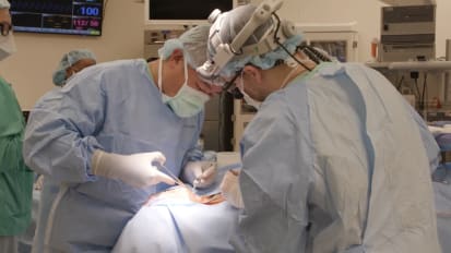
Oliver F. Adunka, MD, FACS, and Matthew Old, MD, perform a pediatric translabyrinthine procedure to the cerebello-pontine angle to remove a tumor. Dr. Adunka and Dr. Old removed the lesion that had caused hearing loss and facial nerve paralysis in the patient.
Related Presenters


Related Videos